 Back
The Integumentary System, Bones, and Joints: ANP Study Guide
Study Guide - Smart Notes
 Tailored notes based on your materials, expanded with key definitions, examples, and context.
The Integumentary System
Introduction to the Integumentary System
The integumentary system is composed of the skin and its accessory structures, including hair, nails, and glands. It serves as the body's first line of defense and plays a vital role in protection, sensation, thermoregulation, and metabolic functions.
Skin Structure
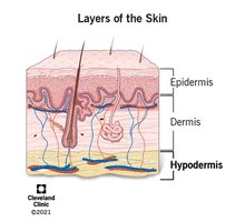
Epidermis: The outermost layer, composed of stratified squamous epithelial cells. It is avascular (contains no blood vessels).
Dermis: The middle layer, made of strong, flexible connective tissue. Contains nerves, blood vessels, hair follicles, and glands.
Hypodermis (Subcutaneous Layer): Not technically part of the skin, but anchors skin to underlying tissues and organs. Composed mainly of adipose tissue.
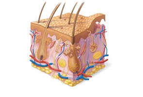
The Epidermis
The epidermis consists of several layers (strata), with the number of layers depending on the region of the body:
Stratum corneum (outermost, dead cells)
Stratum lucidum (only in thick skin, e.g., palms, soles)
Stratum granulosum
Stratum spinosum
Stratum basale (deepest, mitotically active layer)
Keratinocytes, melanocytes, dendritic cells, and tactile cells are the main cell types found in the epidermis.
The Dermis
The dermis is divided into two layers:
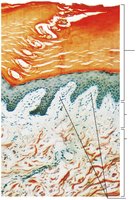
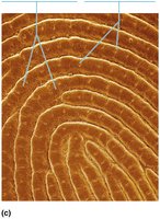
Papillary layer: Areolar connective tissue with dermal papillae that project into the epidermis, forming friction ridges (fingerprints).
Reticular layer: Dense irregular connective tissue, providing strength and elasticity. Contains most of the skin's structures, including glands and hair follicles.
Skin Pigments
Skin color is determined by three main pigments:
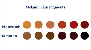
Melanin: Produced by melanocytes; two types are pheomelanin (yellow-red) and eumelanin (brown-black).
Carotene: Yellow-orange pigment stored in the stratum corneum and adipose tissue.
Hemoglobin: Red pigment in blood, visible through the skin.
The Hypodermis
The hypodermis (subcutaneous layer) is composed mainly of adipose tissue and serves as insulation, energy storage, and shock absorption. It also contains blood vessels and sometimes bursa (fluid-filled sacs).
Accessory Structures of the Skin
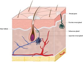
Hair: Produced by hair follicles; associated with arrector pili muscles and sebaceous glands.
Nails: Modifications of the epidermis, composed of hard keratin.
Glands:
Sudoriferous (Sweat) glands: Eccrine (widely distributed, thermoregulation) and apocrine (armpits, anogenital areas, function at puberty).
Sebaceous (Oil) glands: Secrete sebum, associated with hair follicles.
Ceruminous glands: Modified sweat glands in the ear canal.

Functions of the Skin
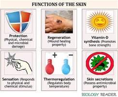
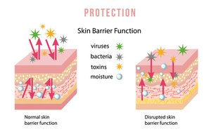
Protection: Physical, chemical, and biological barrier against pathogens, UV radiation, and dehydration.
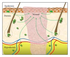
Regeneration: Stem cells in the stratum basale allow for repair and renewal.
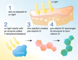
Synthesis of Vitamin D: UV light converts precursors in the skin to vitamin D, essential for calcium absorption.
Cutaneous Sensations: Sensory receptors detect touch, pressure, pain, and temperature.
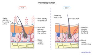
Thermoregulation: Sweat production and blood flow adjustments help regulate body temperature.
Excretion and Absorption: Minor role in excreting wastes and absorbing substances.
Protection Mechanisms
Keratin: Waterproofs and protects against abrasion.
Lipids: Released by lamellar granules, prevent water loss.
Sebum: Hydrates and has antimicrobial properties.
Acidic Sweat: Inhibits microbial growth.
Melanin: Absorbs UV radiation.
Macrophages: Immune defense via phagocytosis.
Regeneration
Stem cells in the stratum basale continually divide to replace lost or damaged cells. Dead cells are shed from the surface, revealing new cells beneath.
Synthesis of Vitamin D
UV rays from sunlight convert 7-dehydrocholesterol in the skin to vitamin D3, which is then modified in the liver and kidneys to form active vitamin D (calcitriol). Calcitriol is essential for calcium absorption in the gut.
Thermoregulation
The skin regulates body temperature through sweat production and changes in blood flow to the dermis. Vasodilation increases heat loss, while vasoconstriction conserves heat.
Age-Associated Changes
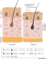
Wrinkles and decreased elasticity
Dehydration and cracking
Decreased sweat production
Loss of melanocytes (gray hair, atypical pigmentation)
Loss of subcutaneous fat and thinning of skin
Brittle nails
Bones and Skeletal Tissues
Types of Cartilage
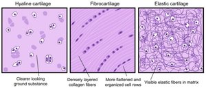
Hyaline cartilage: Most abundant, provides support and flexibility (e.g., articular surfaces, costal cartilages).
Elastic cartilage: Contains elastic fibers, found in the external ear and epiglottis.
Fibrocartilage: Thick collagen fibers, high tensile strength (e.g., intervertebral discs, menisci).
Growth of Cartilage
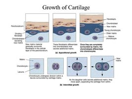
Appositional growth: New matrix is secreted on the surface by chondroblasts in the perichondrium.
Interstitial growth: Chondrocytes divide and secrete new matrix from within the cartilage.
Calcification of cartilage occurs during normal bone growth and aging, but calcified cartilage is not bone.
Classification of Bones
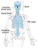
Axial skeleton: Skull, vertebral column, rib cage (long axis of body).
Appendicular skeleton: Limbs and girdles attaching them to the axial skeleton.
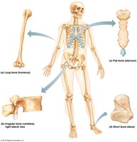
Long bones: Longer than wide (e.g., femur, humerus).
Short bones: Cube-shaped (e.g., carpals, tarsals); sesamoid bones form within tendons (e.g., patella).
Flat bones: Thin, flat, slightly curved (e.g., sternum, skull bones).
Irregular bones: Complicated shapes (e.g., vertebrae, hip bones).
Structure of a Long Bone
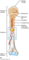
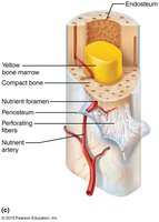
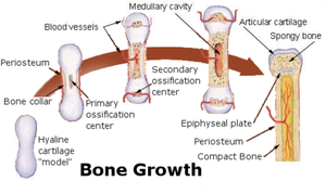
Diaphysis: Shaft, composed of compact bone surrounding the medullary cavity (yellow marrow in adults).
Epiphyses: Bone ends, compact bone externally, spongy bone internally, covered by articular cartilage.
Epiphyseal line: Remnant of the growth plate.
Membranes: Periosteum (outer, fibrous and osteogenic layers) and endosteum (internal, lines medullary cavity).
Bone Markings
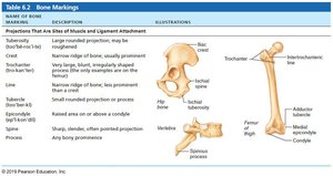
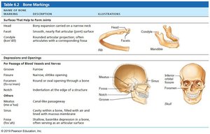
Bone markings are projections, depressions, and openings that serve as sites for muscle, ligament, and tendon attachment, or as passages for nerves and blood vessels.
Name | Description | Example |
|---|---|---|
Crest | Narrow ridge of bone | Iliac crest |
Condyle | Rounded articular projection | Mandibular condyle |
Foramen | Round or oval opening | Foramen magnum |
Meatus | Canal-like passageway | External acoustic meatus |
Fossa | Shallow depression | Mandibular fossa |
Spine | Sharp, slender projection | Ischial spine |
Tuberosity | Large, rounded projection | Tibial tuberosity |
Head | Bony expansion on a narrow neck | Femoral head |
Microscopic Anatomy of Bone
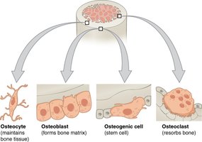
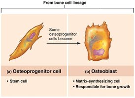
Osteogenic cells: Stem cells in periosteum and endosteum.
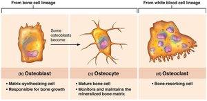
Osteoblasts: Bone-forming cells, secrete osteoid (unmineralized bone matrix).
Osteocytes: Mature bone cells, maintain bone matrix.
Bone-lining cells: Flat cells on bone surfaces, help maintain matrix.
Osteoclasts: Bone-resorbing cells, break down bone matrix.
Bone Development (Ossification)
Endochondral ossification: Bone forms by replacing hyaline cartilage (most bones).
Intramembranous ossification: Bone develops from fibrous membrane (e.g., flat bones of skull, clavicle).
Joints (Articulations)
Classification of Joints
Structural classification: Based on material binding bones and presence of joint cavity (fibrous, cartilaginous, synovial).
Functional classification: Based on movement allowed (synarthrosis—immovable, amphiarthrosis—slightly movable, diarthrosis—freely movable).
Fibrous Joints
Sutures: Rigid, interlocking joints of the skull.
Syndesmoses: Bones connected by ligaments (e.g., distal tibiofibular joint).
Gomphoses: Peg-in-socket joints (e.g., teeth in alveolar sockets).
Cartilaginous Joints
Synchondroses: Bones united by hyaline cartilage (e.g., epiphyseal plates, first rib and sternum).
Symphyses: Bones united by fibrocartilage (e.g., intervertebral discs, pubic symphysis).
Synovial Joints
Synovial joints are freely movable and characterized by a joint cavity filled with synovial fluid. They have articular cartilage, a joint capsule, synovial membrane, and supporting ligaments.
Fatty pads: Cushioning between capsule and bone.
Articular discs (menisci): Improve fit, stabilize joint, reduce wear.
Bursae and tendon sheaths: Reduce friction where structures rub together.
Types of Synovial Joints
Plane: Gliding movements (e.g., intercarpal joints).
Hinge: Uniaxial movement (e.g., elbow, knee).
Pivot: Rotation (e.g., proximal radioulnar joint).
Condylar: Biaxial movement (e.g., knuckle joints).
Saddle: Biaxial movement (e.g., thumb joint).
Ball-and-socket: Multiaxial movement (e.g., shoulder, hip).
Movements Allowed by Synovial Joints
Gliding: Flat surfaces slide past each other.
Angular: Flexion, extension, hyperextension, abduction, adduction, circumduction.
Rotation: Turning of a bone around its own long axis.
Special movements: Supination, pronation, dorsiflexion, plantar flexion, inversion, eversion, protraction, retraction, elevation, depression, opposition.
Factors Influencing Joint Stability
Shape of articular surfaces
Number and location of ligaments
Muscle tone (most important)
Selected Synovial Joints
Shoulder joint: Most freely movable, but easily dislocated.
Elbow joint: Hinge joint stabilized by collateral ligaments.
Hip joint: Ball-and-socket, very stable due to deep socket and strong ligaments.
Knee joint: Largest, most complex, susceptible to injury.
Aging and Joints
Decreased synovial fluid production
Thinning of articular cartilage
Loss of ligament flexibility
