 Back
The Integumentary System II: Skin Color, Glands, and Homeostatic Imbalances
Study Guide - Smart Notes
 Tailored notes based on your materials, expanded with key definitions, examples, and context.
The Integumentary System II
Skin Color
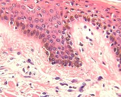
The color of human skin is determined by a combination of genetic, physiological, and environmental factors. The primary contributors to skin color are the pigments melanin, carotene, and hemoglobin. All humans have roughly equal numbers of melanocytes, but the amount and type of melanin produced varies, leading to differences in skin tone. Freckles and pigmented moles are localized accumulations of melanin.
Melanin: A pigment ranging from yellow to black, produced by melanocytes. It protects against ultraviolet (UV) radiation damage.
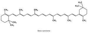
Carotene: A yellow-orange pigment obtained from the diet, especially from vegetables like carrots and sweet potatoes. It accumulates in the stratum corneum and fatty tissue of the hypodermis.
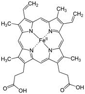
Hemoglobin: The red pigment in red blood cells that gives a pinkish hue to fair skin due to the transparency of the epidermis.
Tan: Increased melanin production in response to UV exposure.
Freckles and Pigmented Moles: Localized concentrations of melanin.




Homeostatic Imbalances of Skin Color
Alterations in skin color can indicate underlying health conditions or genetic disorders. These changes are often due to abnormal pigment production, blood flow, or the presence of foreign substances in the skin.
Cyanosis: A bluish discoloration of the skin due to low oxygen levels in the blood.
Albinism: A genetic disorder characterized by a lack of melanin production, resulting in very pale skin, hair, and eyes.
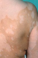
Vitiligo: An autoimmune condition causing loss of melanocytes in patches, leading to depigmented areas of skin.
Redness (Erythema): Increased blood flow to the skin, often due to inflammation, fever, or emotional responses.
Jaundice: Yellowing of the skin and eyes due to bile pigment accumulation, often indicating liver dysfunction.
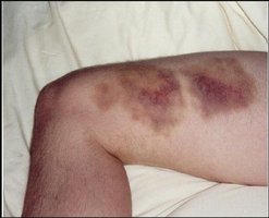
Bruises (Hematomas): Localized collections of clotted blood beneath the skin, resulting from trauma.
Sweat Glands
The human body contains approximately 2.5 million sweat glands, which play a crucial role in thermoregulation and excretion. There are several types of sweat glands, each with distinct functions and secretions.
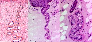
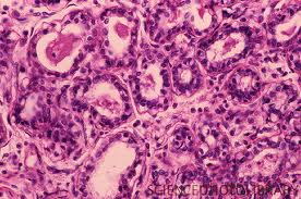
Eccrine Sweat Glands: The most numerous type, found on the palms, soles, and forehead. They secrete a watery sweat that helps cool the body.
Apocrine Sweat Glands: Located mainly in the axillary and genital regions. Their secretion is more viscous and can develop odor when decomposed by skin bacteria.
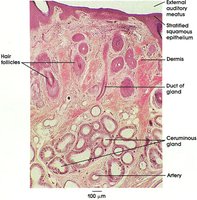
Ceruminous Glands: Modified sweat glands in the ear canal that produce earwax (cerumen).
Mammary Glands: Specialized glands that secrete milk.
Eccrine Sweat Glands
Eccrine glands are distributed widely across the body and are especially abundant on the palms, soles, and forehead. Their primary function is thermoregulation through the secretion of sweat.
Secretion: Sweat is composed of 99% water, some salts, dermicidin (an antimicrobial protein), drugs, and metabolic wastes such as urea, uric acid, and ammonia.
pH: Sweat is slightly acidic, with a pH of 4-6.
Apocrine Sweat Glands
Apocrine glands are confined to the axillary and genital regions. Their ducts empty into hair follicles, and their secretions contain fatty substances and proteins, making them more viscous than eccrine sweat. The secretion is initially odorless but can develop a characteristic body odor (BO) when decomposed by skin bacteria. The exact function of apocrine glands in humans is not fully understood but may be related to sexual scent communication, as in other animals.
Sebaceous (Oil) Glands
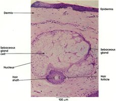
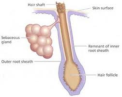
Sebaceous glands are found throughout the skin except on the palms and soles. They secrete sebum, an oily substance that lubricates and protects hair and skin and has bactericidal properties.
Sebum: Oily lipids that help prevent hair and skin from drying out and provide a barrier against some bacteria.
Distribution: Absent on the palms and soles but present elsewhere, often associated with hair follicles.
Homeostatic Imbalances of Glands
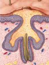
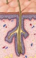
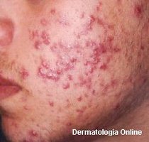
Disorders of the sebaceous glands can lead to common skin conditions such as whiteheads, blackheads, and acne.
Whiteheads: Blocked sebaceous glands filled with sebum and dead cells.
Blackheads: Similar to whiteheads, but the material in the blocked gland is oxidized, turning it black.
Acne: Inflammation of sebaceous glands, often due to bacterial infection, which can lead to scarring if severe.
Additional info: The integumentary system is essential for protection, sensation, thermoregulation, and metabolic functions. Disorders of pigmentation and glandular function can serve as important clinical indicators of systemic health.
