 Back
BackThe Integumentary System: Structure and Function
Study Guide - Smart Notes
 Tailored notes based on your materials, expanded with key definitions, examples, and context.
The Integumentary System
Overview
The integumentary system is the body's largest organ system, primarily composed of the skin and its accessory structures. It serves as a protective barrier, regulates body temperature, and plays a vital role in homeostasis.
Skin Structure
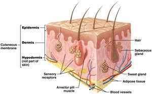
Main Layers of the Skin
Epidermis: The superficial layer, consisting of keratinized stratified squamous epithelium. It is avascular and relies on diffusion from the dermis for nutrients and oxygen.
Dermis: Located beneath the epidermis, composed of loose connective tissue and dense irregular connective tissue. It is highly vascular and provides structural support, sensory receptors, and anchors the epidermis.
Hypodermis (Superficial Fascia): Not technically part of the skin, but anchors the skin to underlying structures. Composed of loose connective and adipose tissue, it contains abundant blood vessels.
Cellulite
Definition: Dimpled or "orange peel" appearance of skin due to collagen bands forming around adipose tissue in the hypodermis.
Factors: Genetics, gender, adipose tissue distribution, and age influence its development.
Management: Healthy diet and exercise may minimize appearance, but do not eliminate it entirely.
Functions of the Integumentary System
Protection
Protects underlying tissues from mechanical trauma, pathogens, and environmental hazards.
Sensation
Contains sensory receptors that detect changes in the internal and external environment, critical for homeostasis.
Thermoregulation
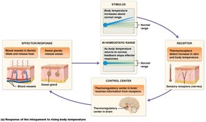
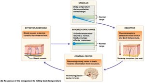
The integumentary system maintains stable internal temperature through negative feedback mechanisms.
When body temperature rises: Thermoreceptors detect the change, the hypothalamus (control center) stimulates sweat production and cutaneous vasodilation, increasing heat loss.
When body temperature falls: Thermoreceptors signal the hypothalamus to trigger vasoconstriction, reducing blood flow to the skin and conserving heat.
Excretion
Eliminates small amounts of waste products and toxins through sweat.
Vitamin D Synthesis
Deep epidermal cells convert a cholesterol-based precursor to cholecalciferol (vitamin D3) upon UV exposure. The liver and kidneys further modify it to calcitriol, the active form of vitamin D, essential for calcium absorption.
The Epidermis
Keratinocytes
Comprise about 95% of the epidermis.
Produce keratin, a tough protein that provides mechanical strength.
Linked by desmosomes for structural integrity.
Keratinocyte Life Cycle
Originate in the stratum basale or spinosum and migrate to the surface, becoming more keratinized and eventually dying.
The journey from the deepest layer to the stratum corneum takes 40–50 days.
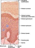
Other Epidermal Cells
Dendritic (Langerhans) cells: Immune cells in the stratum spinosum.
Merkel cells: Sensory receptors for light touch in the stratum basale.
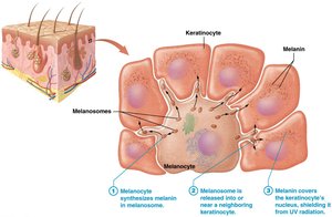
Melanocytes: Produce melanin pigment in the stratum basale.
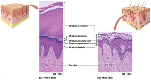
Thick and Thin Skin
Thick skin: Found on palms and soles, contains all five epidermal layers, including a prominent stratum corneum. Lacks hair follicles but has many sweat glands.
Thin skin: Covers most of the body, missing the stratum lucidum, and has thinner layers overall. Contains hair follicles, sweat, and sebaceous glands.
Callus: Localized thickening of the stratum corneum due to repetitive pressure.
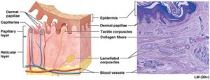
The Dermis
Structure and Function
Highly vascular, provides nutrients to the epidermis, contains sensory receptors, and anchors the epidermis.
Composed of two layers: papillary (loose connective tissue) and reticular (dense irregular connective tissue).
Skin Markings
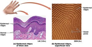
Dermal and Epidermal Ridges
Dermal ridges in thick skin create epidermal ridges (fingerprints), enhancing grip and forming unique patterns.
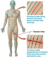
Tension (Cleavage) Lines
Lines formed by the orientation of collagen bundles in the dermis; important for surgical incisions as cuts parallel to these lines heal better.
Skin Pigmentation
Melanin
Produced by melanocytes, melanin protects DNA from UV-induced mutations and determines skin color.
Increased UV exposure stimulates melanin production, resulting in tanning.
Other Pigments
Carotene: Yellow-orange pigment from diet.
Hemoglobin: Red pigment in blood, contributes to skin color based on blood flow.
Clinical Significance of Skin Color
Erythema: Redness due to increased blood flow (e.g., fever, infection).
Pallor: Paleness from decreased blood flow (e.g., cold, shock).
Cyanosis: Bluish tint indicating low oxygen levels in blood.
Accessory Structures
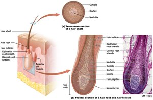
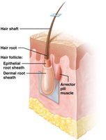
Hair
Composed of a shaft (visible) and root (embedded in skin), both made of keratinized epithelial cells.
The root is surrounded by the hair follicle, which includes epithelial and dermal root sheaths.
Arrector pili muscles cause hair to stand up (goosebumps).
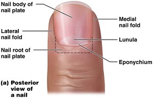
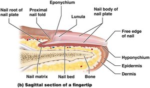
Nails
Hard structures at the ends of digits, composed of keratinized stratified squamous epithelium.
Consist of the nail plate (body and root), nail matrix (growth region), and surrounding folds (eponychium/cuticle).
Primary function is protection and aiding in manipulation of objects.
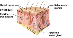
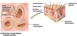
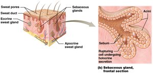
Glands
Sudoriferous (Sweat) Glands: Eccrine (most common, for thermoregulation), apocrine (in axillae, become active after puberty), ceruminous (ear wax), and mammary (milk production).
Sebaceous Glands: Secrete sebum (oily substance) into hair follicles, providing lubrication and antibacterial action. Activity increases after puberty.
Common Skin Pathologies
Acne Vulgaris
Results from accumulation of sebum and dead cells in sebaceous glands, sometimes leading to infection and inflammation.
Most common in adolescents due to increased androgen levels.
Wounds and Skin Cancer
Wounds: Include lacerations, burns, and cancers; disrupt skin integrity.
Skin Cancer: Caused by mutations in DNA, often due to UV exposure, leading to uncontrolled cell division and tumor formation.
