 Back
BackThe Integumentary System: Structure and Function
Study Guide - Smart Notes
 Tailored notes based on your materials, expanded with key definitions, examples, and context.
The Integumentary System
Overview and Functions
The integumentary system is the body's largest organ system, serving as the primary interface between the body and the external environment. It is essential for protection, homeostasis, and sensory perception.
Protection: Acts as a barrier against mechanical injury, pathogens, and harmful substances.
Excretion: Removes metabolic wastes through sweat glands.
Temperature Maintenance: Regulates body temperature via sweat and blood flow.
Insulation and Cushion: Provides thermal insulation and cushions underlying tissues.
Vitamin D3 Synthesis: Initiates synthesis of vitamin D3 when exposed to UV light.
Sensory Detection: Contains receptors for touch, pressure, pain, and temperature.
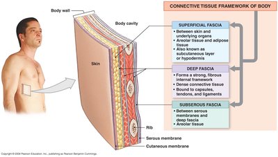
Components of the Integumentary System
Cutaneous Membrane: Composed of the epidermis (superficial epithelial layer) and dermis (deeper connective tissue layer).
Accessory Structures: Includes hair, nails, and glands (sebaceous and sweat glands).
Subcutaneous Layer (Hypodermis): Not technically part of the integumentary system, but separates it from underlying deep fascia. Composed mainly of adipose and areolar tissue, providing insulation and energy storage.
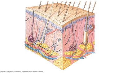
Structure of the Skin
Layers of the Skin
The skin consists of three main layers: the epidermis, dermis, and subcutaneous layer (hypodermis). Each layer has distinct structures and functions.
Epidermis: Outermost, avascular layer composed of stratified squamous epithelium.
Dermis: Middle, vascular layer containing connective tissue, blood vessels, nerves, and accessory structures.
Subcutaneous Layer (Hypodermis): Deepest layer, primarily adipose tissue, not part of the skin proper.
Cells of the Epidermis
Keratinocytes: Most abundant; produce keratin, a protein that provides toughness and water resistance.
Melanocytes: Produce melanin, which protects against UV radiation and determines skin color.
Langerhans Cells: Immune cells that help defend against pathogens.
Merkel Cells: Associated with sensory nerve endings; involved in touch sensation.
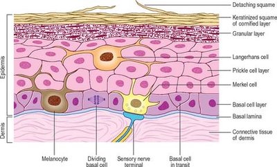
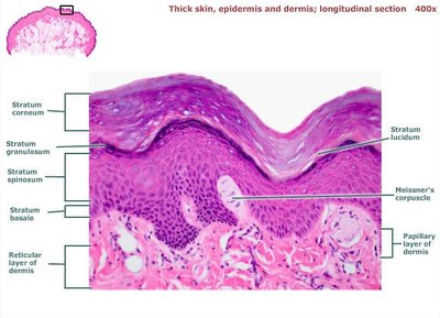
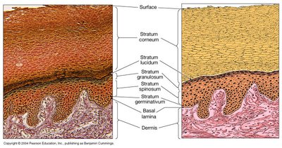
Layers of the Epidermis
The epidermis is organized into distinct layers (strata), each with specialized functions. Thick skin (palms, soles) has five layers; thin skin (most of the body) has four.
Stratum Basale (Germinativum): Deepest layer; single row of dividing cells, contains melanocytes and Merkel cells.
Stratum Spinosum: Several layers of keratinocytes; contains Langerhans cells and desmosomes for strength.
Stratum Granulosum: 3–5 layers; cells contain keratohyalin and lamellated granules, begin to die and lose nuclei.
Stratum Lucidum: Present only in thick skin; thin, clear layer of dead keratinocytes.
Stratum Corneum: Outermost layer; 20–30 layers of dead, keratinized cells, continuously shed and replaced.

Specialized Features of the Epidermis
Thick Skin: Found on palms and soles; contains all five layers, especially prominent stratum lucidum and thick stratum corneum.
Thin Skin: Covers most of the body; lacks stratum lucidum, thinner stratum corneum.
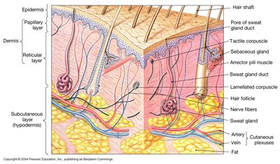
The Dermis
Dermal Layers
The dermis provides structural strength and elasticity to the skin. It is divided into two layers:
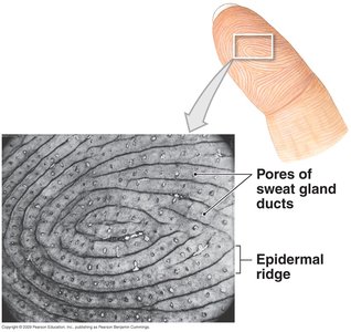
Papillary Layer: Superficial; composed of loose areolar connective tissue, contains capillaries, lymphatics, sensory neurons, and dermal papillae (finger-like projections that form fingerprints).
Reticular Layer: Deeper; dense irregular connective tissue with collagen and elastic fibers, houses sweat and sebaceous glands, hair follicles, and deep pressure receptors (Pacinian corpuscles).
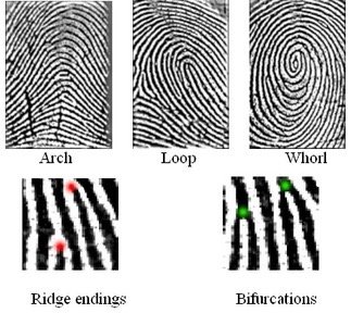
Dermal Papillae and Fingerprints
Dermal papillae interlock with epidermal ridges, increasing surface area for attachment and forming unique fingerprint patterns. These patterns are genetically determined and remain unchanged throughout life.
Cells and Structures in the Dermis
Fibroblasts: Produce collagen and elastic fibers.
Adipocytes: Store fat, especially in the hypodermis.
Macrophages: Immune defense.
Blood Vessels: Supply nutrients, regulate temperature.
Nerve Endings: Detect touch, pressure, pain, and temperature.
Skin Color
Determinants of Skin Color
Blood Supply: Oxygenated blood imparts a reddish hue; reduced oxygen causes cyanosis (bluish color).
Carotene: Yellow-orange pigment from diet, accumulates in stratum corneum and hypodermis.
Melanin: Brown-black pigment produced by melanocytes; amount (not number of cells) determines skin color.
Abnormal Skin Colors: Flushed (fever, hypertension), pale, jaundice (yellow, liver dysfunction), cyanosis (blue, low oxygen).
Accessory Structures
Nails
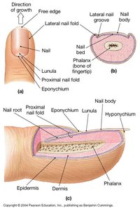
Nails are protective coverings on the dorsal surface of fingers and toes. They are composed of hard keratin.
Nail Body: Visible part covering the nail bed.
Nail Matrix: Site of nail production.
Eponychium (Cuticle): Overlies the root.
Lunula: Crescent-shaped area at the base.
Free Edge: Extends beyond the finger or toe.
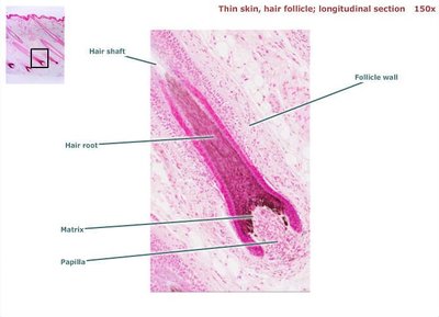
Hair
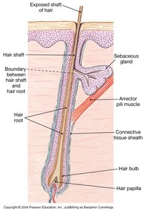
Hair is composed of dead, keratinized cells and serves protective and sensory functions.
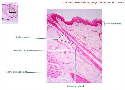
Hair Follicle: Invagination of the epidermis into the dermis, surrounds the hair root.
Hair Shaft: Portion above the skin surface.
Hair Root: Portion below the surface, ends in the hair bulb.
Hair Papilla: Contains blood vessels that nourish the growing hair.
Arrector Pili Muscle: Smooth muscle that causes hair to stand (goosebumps).
Cutaneous Glands
Sebaceous (Oil) Glands
Sebaceous glands are holocrine glands that secrete sebum (oil and dead cells) into hair follicles or directly onto the skin. Sebum lubricates and waterproofs the skin and hair. These glands are absent on the palms and soles.
Functions: Prevents drying, inhibits bacterial growth, can contribute to acne if blocked.
Sudoriferous (Sweat) Glands
Apocrine Sweat Glands: Located in axilla and genital areas; begin functioning at puberty; produce viscous, protein-rich, odorous secretion.
Merocrine (Eccrine) Sweat Glands: Distributed over most of the body; produce watery sweat for thermoregulation; excrete water, salts, and urea.
Summary Table: Layers of the Epidermis
Layer | Location | Key Features |
|---|---|---|
Stratum Corneum | Surface | 20–30 layers of dead, keratinized cells; barrier function |
Stratum Lucidum | Thick skin only | Clear, dead keratinocytes |
Stratum Granulosum | Middle | 3–5 layers; keratohyalin and lamellated granules |
Stratum Spinosum | Above basale | Several layers; desmosomes; Langerhans cells |
Stratum Basale | Deepest | Single row; mitotic cells; melanocytes; Merkel cells |
