 Back
BackThe Integumentary System: Structure and Function
Study Guide - Smart Notes
 Tailored notes based on your materials, expanded with key definitions, examples, and context.
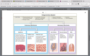
The Integumentary System
Overview and Functions
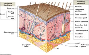
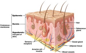
The integumentary system is the body's largest organ system, consisting of the skin and its accessory structures. It serves as a protective barrier, regulates temperature, and provides sensory information. The system is composed of three main layers: the epidermis, dermis, and subcutaneous (hypodermis) layer, along with accessory structures such as hair, nails, and exocrine glands.
Protection: Shields underlying tissues from abrasion, fluid loss, and pathogens.
Excretion: Removes salts, water, and organic wastes via sweat and oil glands.
Temperature Homeostasis: Maintains body temperature through insulation, vessel dilation/constriction, and sweating.
Vitamin D3 Synthesis: Converts precursor molecules to vitamin D3 in response to sunlight.
Nutrient Storage: Stores lipids in the subcutaneous layer.
Sensation: Detects touch, pressure, pain, and temperature.
Immunity: Provides immune defense via specialized cells.
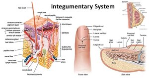
Layers of the Integumentary System
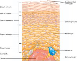
Epidermis
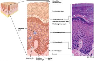
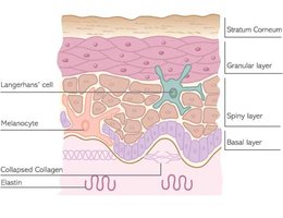
The epidermis is the outermost layer of the skin, providing a physical barrier against environmental hazards. It is composed of keratinized stratified squamous epithelium and is avascular. The main cell type is the keratinocyte, which produces keratin for strength and waterproofing.
Thin Skin: Contains four sub-layers; covers most of the body.
Thick Skin: Contains five sub-layers; found on palms and soles.
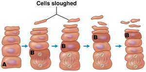
Cell Life Cycle: Cells divide in the deepest layers and are pushed upward, dying as they move toward the surface.
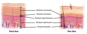
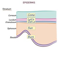
Layers of the Epidermis (Deep to Superficial)
Stratum Basale: Single layer of stem cells; contains melanocytes and Merkel cells; site of cell division and vitamin D synthesis.
Stratum Spinosum: Thickest layer; contains keratinocytes and dendritic (Langerhans) cells; still metabolically active.
Stratum Granulosum: Cells fill with keratin and granules, then die; provides waterproofing.
Stratum Lucidum: Clear, dead keratinocytes; found only in thick skin.
Stratum Corneum: 15-30 layers of dead, keratinized cells; tightly interconnected by desmosomes; sloughed off regularly.

Keratinocyte Life Cycle
Mitosis: Occurs in stratum basale and spinosum.
Migration: Cells are pushed upward, taking 7-10 days to reach the stratum corneum, where they remain for ~2 weeks before shedding.
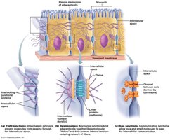
Desmosomes: Permanent connections between cells, allowing shedding in sheets.


Other Cells of the Epidermis
Dendritic (Langerhans) Cells: Immune phagocytes in the stratum spinosum; eat pathogens.
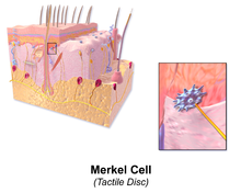
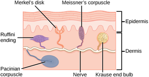
Merkel Cells: Sensory receptor cells in the stratum basale; detect light touch, shapes, and textures.
Melanocytes: Pigment cells in the stratum basale; produce melanin.
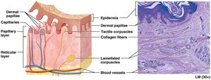
Dermis
Structure and Function
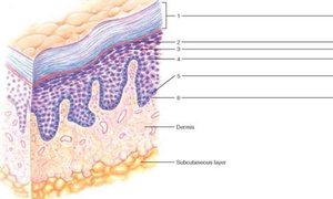
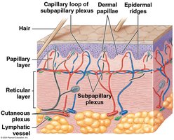
The dermis is the tissue layer beneath the epidermis, providing structural support and housing accessory structures. It contains blood vessels, nerves, and connective tissue, and is divided into two sub-layers: the papillary layer and the reticular layer.
Papillary Layer: Thinner, areolar connective tissue; anchors dermis to epidermis; contains dermal papillae and tactile (Meissner) corpuscles for light touch.
Reticular Layer: Thicker, dense irregular connective tissue; contains collagen and elastic fibers, blood vessels, sweat glands, hair follicles, and lamellated (Pacinian) corpuscles for pressure and vibration.
Functions: Provides blood supply for the epidermis, sensory receptors, and anchors the epidermis.


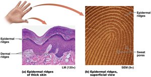
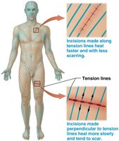
Tension Lines
Tension Lines (Cleavage Lines): Created by prominent dermal papillae and thick collagen bundles; result in epidermal ridges (fingerprints).
Clinical Importance: Incisions made along tension lines heal faster and with less scarring.
Dermal Blood Supply
Subpapillary Plexus: Capillary network supplying the dermis and epidermis.
Cutaneous Plexus: Deeper blood vessels in the dermis and hypodermis.
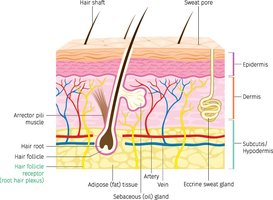
Subcutaneous Layer (Hypodermis)
Structure and Function
The subcutaneous layer, or hypodermis, is not technically part of the skin but connects the dermis to underlying tissues. It is composed mainly of adipose and areolar connective tissue, providing insulation, cushioning, and energy storage.
Connection: Links skin to muscles and bones below.
Nerve & Blood Vessels: Contains large vessels and nerves.
Insulation & Cushioning: Fat provides temperature regulation and protection.
Energy Homeostasis: Stores fat for energy.
Skin Color: Pigmentation and Circulation
Melanocytes and Melanin
Skin color is determined by epidermal pigmentation and dermal circulation. Melanocytes in the stratum basale produce melanin, which is transferred to keratinocytes. The number of melanocytes is similar across individuals, but the amount and type of melanin produced varies.
Melanin: Provides UV protection; two types—red-yellow (pheomelanin) and brown-black (eumelanin).
Carotene: Contributes to skin color; stored in the epidermis and subcutaneous fat.
Melanosomes: Vesicles that package melanin and transfer it to keratinocytes.
UV Exposure: Accelerates melanin production.
HTML Table: Layers and Functions of the Integumentary System
Layer | Structure | Function |
|---|---|---|
Epidermis | Keratinized stratified squamous epithelium | Physical barrier, prevents fluid loss, abrasion resistance |
Dermis - Papillary Layer | Areolar connective tissue, dermal papillae | Anchors epidermis, provides blood supply, light touch sensation |
Dermis - Reticular Layer | Dense irregular connective tissue, collagen & elastic fibers | Strength, elasticity, houses accessory structures |
Subcutaneous Layer (Hypodermis) | Adipose & areolar connective tissue | Insulation, energy storage, connection to underlying tissues |
Key Terms and Definitions
Keratinocyte: Main cell type in the epidermis, produces keratin.
Melanocyte: Cell that produces melanin pigment.
Desmosome: Cell junction providing strong adhesion between cells.
Dermal Papillae: Projections of the dermis that anchor the epidermis and increase surface area for diffusion.
Tactile (Meissner) Corpuscle: Sensory receptor for light touch.
Lamellated (Pacinian) Corpuscle: Sensory receptor for pressure and vibration.
Equations and Formulas
Insensible Perspiration: Water diffusion and evaporation across the stratum corneum.
Vitamin D3 Synthesis:
Short Comparison: Thick vs. Thin Skin
Feature | Thick Skin | Thin Skin |
|---|---|---|
Location | Palms, soles | Most of body |
Layers | 5 (includes stratum lucidum) | 4 (no stratum lucidum) |
Hair | Absent | Present |
Sweat Glands | More numerous | Less numerous |
Example: Clinical Application of Tension Lines
Surgeons make incisions along tension lines to minimize scarring and promote faster healing. Incisions perpendicular to tension lines heal more slowly and tend to scar.
Summary
The integumentary system is essential for protection, sensation, temperature regulation, and immune defense. Understanding its structure and function is fundamental for anatomy and physiology students.
