 Back
BackThe Integumentary System: Structure and Function
Study Guide - Smart Notes
 Tailored notes based on your materials, expanded with key definitions, examples, and context.
The Integumentary System
Overview
The integumentary system is composed of the skin and its accessory structures, including hair, nails, sweat glands, sebaceous glands, and arrector pili muscles. This system serves as the body's primary barrier against the external environment and plays a vital role in protection, sensation, thermoregulation, and metabolic functions.
Skin: The largest organ of the body, consisting of two main layers—the epidermis and dermis.
Accessory Structures: Hair, nails, sweat glands, sebaceous (oil) glands, and arrector pili muscles.
Hypodermis (Subcutaneous Tissue): A layer of adipose tissue beneath the dermis, not considered part of the skin but important for insulation and energy storage.
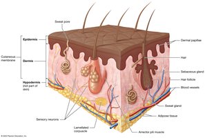
Skin Structure
Main Components
Epidermis: The superficial layer, composed of keratinized stratified squamous epithelium. Provides a waterproof barrier and creates our skin tone.
Dermis: The deeper layer, made of dense irregular connective tissue. Contains blood vessels, nerves, hair follicles, and glands.
Hypodermis: Also called subcutaneous tissue, primarily adipose tissue. Functions in insulation and energy storage but is not technically part of the skin.
The Epidermis
Cell Types and Layers
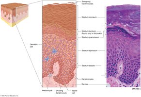
The epidermis is primarily composed of keratinocytes, which produce keratin, a protein that strengthens the skin. Other cell types include melanocytes (produce pigment), dendritic cells (immune function), and tactile cells (sensory function).
Keratinocytes: Main cell type, responsible for producing keratin.
Melanocytes: Produce melanin, the pigment responsible for skin color.
Dendritic (Langerhans) Cells: Immune cells that help detect pathogens.
Tactile (Merkel) Cells: Sensory receptors for touch.
Layers (Strata) of the Epidermis
The epidermis is organized into distinct layers, especially prominent in thick skin (palms, soles):
Stratum basale (deepest)
Stratum spinosum
Stratum granulosum
Stratum lucidum (only in thick skin)
Stratum corneum (most superficial)
Mnemonic for Epidermal Layers
Brilliant – Stratum Basale
Studying – Stratum Spinosum
Gives – Stratum Granulosum
Loads of – Stratum Lucidum
Confidence – Stratum Corneum
Note: Stratum lucidum is only present in thick skin (palms and soles).
Thick vs. Thin Skin
Comparison
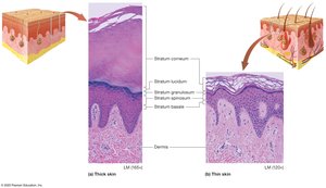
Thick skin is found on the palms of the hands and soles of the feet, while thin skin covers the rest of the body. The main differences are the presence of the stratum lucidum and the thickness of the epidermal layers.
Thick Skin: Contains all five strata, including stratum lucidum; no hair follicles or sebaceous glands.
Thin Skin: Lacks stratum lucidum; contains hair follicles and sebaceous glands.
Accessory Structures of the Skin
Hair
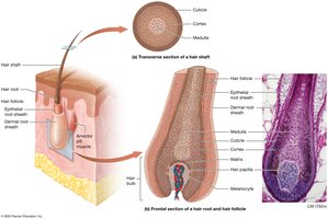
Hair is a filamentous structure composed of dead, keratinized cells. It serves protective, sensory, and thermoregulatory functions.
Hair Shaft: The visible part above the skin surface.
Hair Root: The portion below the skin surface, within the follicle.
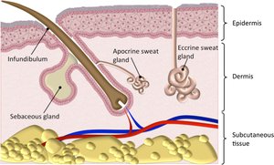
Hair Follicle: The structure that produces hair, extending from the epidermis into the dermis.
Arrector Pili Muscle: Smooth muscle attached to hair follicles; contraction causes "goosebumps."
Glands
The skin contains several types of exocrine glands:
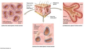
Sweat (Sudoriferous) Glands:
Eccrine Glands: Widely distributed; secrete watery sweat for thermoregulation.
Apocrine Glands: Found in axillary and genital areas; secrete a thicker, milky fluid; become active at puberty.
Sebaceous (Oil) Glands: Secrete sebum into hair follicles; lubricates and waterproofs skin and hair.
Summary Table: Layers and Structures of the Skin
Layer | Main Tissue Type | Key Structures | Functions |
|---|---|---|---|
Epidermis | Keratinized stratified squamous epithelium | Keratinocytes, melanocytes, dendritic cells, tactile cells | Protection, waterproofing, UV protection, sensation |
Dermis | Dense irregular connective tissue | Blood vessels, nerves, hair follicles, glands | Nourishment, sensation, thermoregulation, support |
Hypodermis | Adipose tissue | Fat cells, larger blood vessels | Insulation, energy storage, shock absorption |
Key Terms and Concepts
Keratin: A tough, fibrous protein that provides strength and waterproofing to the skin.
Stratum Basale: The deepest layer of the epidermis; site of cell division.
Stratum Corneum: The outermost layer; consists of dead, keratinized cells.
Sebum: Oily secretion from sebaceous glands; lubricates skin and hair.
Thermoregulation: The process of maintaining a stable internal body temperature, aided by sweat glands and blood vessels in the skin.
Additional info: The integumentary system also plays roles in vitamin D synthesis, immune defense, and excretion of small amounts of waste through sweat.
