 Back
BackThe Integumentary System: Structure and Function
Study Guide - Smart Notes
 Tailored notes based on your materials, expanded with key definitions, examples, and context.
The Integumentary System
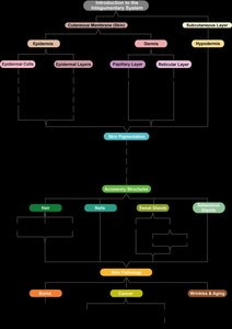
Introduction to the Integumentary System
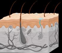
The integumentary system is an organ system composed of the skin (cutaneous membrane), hair, nails, glands, and sensory receptors. It serves as the body's primary barrier to the external environment and plays a vital role in protection, sensation, thermoregulation, and communication.
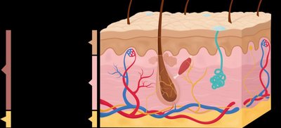
Cutaneous Membrane (Skin): Consists of two main layers—the epidermis (outer epithelial layer) and the dermis (deeper connective tissue layer). Beneath the skin lies the hypodermis (subcutaneous layer), which is not technically part of the skin but anchors it to underlying tissues.
Accessory Structures: Includes hair, nails, and glands (sweat and sebaceous glands).
Functions of the Integumentary System
Protection: Acts as a barrier against mechanical injury, chemicals, UV radiation, and pathogens.
Thermoregulation: Maintains stable internal body temperature through mechanisms such as sweating and blood vessel regulation.
Sensation: Contains sensory receptors for touch, pain, temperature, and pressure.
Communication: Facilitates expressive communication and emotions (e.g., blushing, sweating).
Other Functions: Synthesis of vitamin D, excretion of wastes via sweat, and storage of lipids.
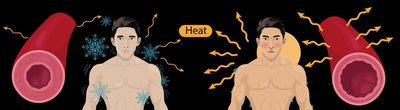
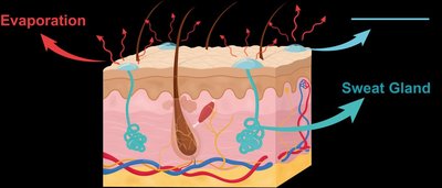
Thermoregulation
Mechanisms of Thermoregulation
The integumentary system maintains homeostasis by regulating body temperature through two main mechanisms: vasoconstriction/vasodilation and sweating.
Vasoconstriction: Blood vessels in the dermis constrict (decrease in diameter) when the body is cold, reducing blood flow to the skin and minimizing heat loss.
Vasodilation: Blood vessels dilate (increase in diameter) when the body is hot, increasing blood flow to the skin and facilitating heat loss.
Sweating: Sweat glands secrete a water-based solution onto the skin surface. As sweat evaporates, it cools the body.
The Epidermis
Cell Types in the Epidermis
The epidermis is composed of stratified squamous epithelial tissue and contains four main cell types:
Keratinocytes: Most abundant cell type; produce keratin, a tough, water-resistant protein that provides mechanical strength and protection.
Melanocytes: Produce melanin, a pigment that protects skin from UV damage.
Dendritic Cells (Langerhans cells): Immune cells that help initiate immune responses and protect against pathogens.
Tactile Epithelial Cells (Merkel cells): Specialized for touch sensation; work with nerve endings to detect mechanical stimuli.
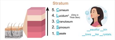
Layers of the Epidermis
The epidermis is organized into distinct layers (strata), each with specialized functions. The number of layers varies between thin and thick skin.
Stratum Basale: Deepest layer; a single row of proliferating cells (keratinocytes, melanocytes, tactile cells). Responsible for generating new epidermal cells.
Stratum Spinosum: Several layers of keratinocytes; contains dendritic cells for immunity. Thickest layer in thin skin.
Stratum Granulosum: Keratinocytes stop dividing, begin to harden, and fill with keratin (keratinization). Cells start to die and lose organelles.
Stratum Lucidum: Present only in thick skin (palms, soles); composed of dead, transparent cells lacking organelles.
Stratum Corneum: Outermost layer; dead, flattened keratinocytes filled with keratin and glycolipids, providing a water-resistant barrier. Cells are regularly shed and replaced.
Thin vs. Thick Skin
Feature | Thin Skin | Thick Skin |
|---|---|---|
Location | Most of the body | Palms of hands, soles of feet |
Stratum Lucidum | Absent | Present |
Hair Follicles & Oil Glands | Present | Absent |
Sweat Glands | Fewer | More numerous |
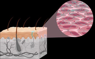
The Dermis
Structure and Layers of the Dermis
The dermis is the second layer of the skin, lying deep to the epidermis. It is composed of two layers:
Papillary Layer: Superficial layer made of loose areolar connective tissue. Contains dermal papillae (projections that indent the epidermis), capillaries, lymphatic vessels, and Meissner corpuscles (touch receptors). Dermal ridges in thick skin enhance grip and form fingerprints.
Reticular Layer: Deeper layer made of dense irregular connective tissue. Contains sweat and oil glands, hair roots, and Pacinian corpuscles (pressure receptors). Collagen and elastic fibers provide strength and flexibility. Cleavage (tension) lines are formed by parallel arrangements of collagen fibers and are important in surgical incisions for optimal healing.
The Hypodermis (Subcutaneous Layer)
Structure and Function
The hypodermis lies deep to the dermis and is not technically part of the skin. It is composed mainly of adipose tissue and some areolar connective tissue.
Anchors the skin to underlying tissues (such as muscle).
Acts as a shock absorber to protect internal organs.
Reduces heat loss by providing insulation.
Serves as an energy reserve due to its fat content.

Summary Table: Layers of the Skin
Layer | Main Tissue | Key Features |
|---|---|---|
Epidermis | Stratified squamous epithelium | Protection, keratinocytes, multiple strata |
Dermis (Papillary) | Areolar connective tissue | Dermal papillae, capillaries, touch receptors |
Dermis (Reticular) | Dense irregular connective tissue | Collagen/elastic fibers, glands, hair roots |
Hypodermis | Adipose & areolar tissue | Anchoring, insulation, energy storage |
Additional info: The integumentary system is essential for maintaining homeostasis, protecting against environmental hazards, and supporting sensory and metabolic functions. Disorders of the skin can impact overall health and are often indicators of systemic disease.
