 Back
The Integumentary System: Structure and Function
Study Guide - Smart Notes
 Tailored notes based on your materials, expanded with key definitions, examples, and context.
The Integumentary System
Overview of the Integumentary System
The integumentary system is composed of the skin and its accessory structures, including hair, nails, and glands. It serves as the body's primary barrier against the external environment and plays a critical role in protection, sensation, thermoregulation, and metabolic functions.
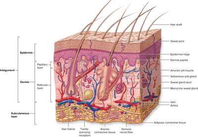
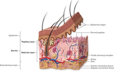
Skin is the largest organ of the body, consisting of three main layers: the epidermis, dermis, and subcutaneous layer (hypodermis).
Accessory structures include hair, nails, and various glands (sebaceous, sweat).
Functions include protection, sensation, temperature regulation, excretion, and synthesis of vitamin D.
Structure of the Skin
Layers of the Skin
The skin is organized into three main layers, each with distinct structures and functions:
Epidermis: The outermost layer, composed of stratified squamous epithelium. It is avascular and provides a waterproof barrier.
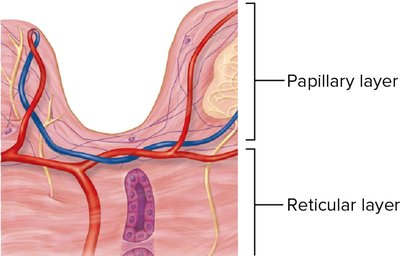
Dermis: The middle layer, made of connective tissue, containing blood vessels, nerves, hair follicles, and glands. It is divided into the papillary and reticular layers.
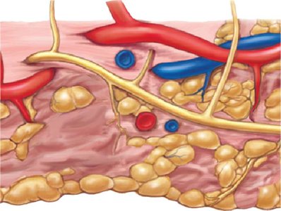
Subcutaneous layer (Hypodermis): The deepest layer, consisting mainly of adipose tissue and areolar connective tissue, providing insulation and cushioning.

Histology of the Epidermis
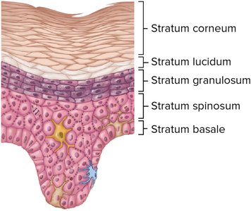
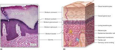
The epidermis is composed of several layers (strata) of keratinocytes, each with specific characteristics:
Stratum basale: The deepest layer, containing stem cells that divide to form new keratinocytes and melanocytes.
Stratum spinosum: Several layers of keratinocytes connected by desmosomes; contains Langerhans cells (immune function).
Stratum granulosum: Keratinocytes begin to die and accumulate keratohyalin granules.
Stratum lucidum: Present only in thick skin (palms, soles); a thin, translucent layer of dead keratinocytes.
Stratum corneum: The outermost layer, consisting of dead, flattened keratinocytes filled with keratin.
Thick vs. Thin Skin
Skin is classified as thick or thin based on the number of epidermal layers and the presence of certain structures:
Thick skin: Found on palms and soles; contains all five strata, including a prominent stratum lucidum; lacks hair follicles and sebaceous glands.
Thin skin: Covers most of the body; lacks stratum lucidum; contains hair follicles, sebaceous, and sweat glands.
Dermis Structure
The dermis provides structural strength and elasticity to the skin. It is divided into two layers:
Papillary layer: Superficial, composed of areolar connective tissue; contains dermal papillae that interlock with epidermal ridges, increasing surface area for nutrient exchange and anchoring.
Reticular layer: Deeper, composed of dense irregular connective tissue; contains collagen and elastic fibers, providing strength and extensibility.
Subcutaneous Layer (Hypodermis)
The subcutaneous layer lies beneath the dermis and is not technically part of the skin but is important for its functions:
Composed mainly of adipose tissue (fat) and areolar connective tissue.
Functions in insulation, energy storage, and shock absorption.
Skin Color and Pigmentation
Melanocytes and Melanin
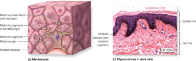
Skin color is primarily determined by the amount and type of melanin produced by melanocytes in the stratum basale:
Melanocytes synthesize melanin, which is transferred to keratinocytes and accumulates above the nucleus to protect DNA from UV radiation.
Variation in skin color is due to the amount, type, and distribution of melanin, not the number of melanocytes.

Other Factors Affecting Skin Color
Carotene: A yellow-orange pigment from diet, accumulates in the stratum corneum and subcutaneous fat.
Hemoglobin: Oxygenated blood imparts a pinkish hue to fair skin.
Surface Features of the Skin
Epidermal Ridges and Fingerprints
Epidermal ridges are formed by the underlying dermal papillae and are responsible for fingerprints:
Increase grip by enhancing friction.
Unique to each individual; patterns include arches, whorls, and loops.
Accessory Structures of the Skin
Nails
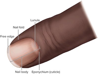
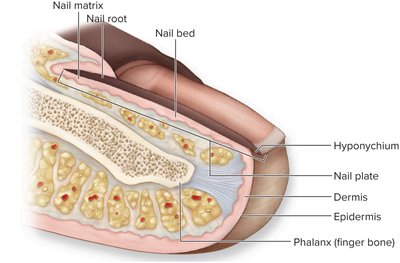
Nails are hard, keratinized structures that protect the distal tips of fingers and toes:
Nail plate: Visible part of the nail.
Nail bed: Skin beneath the nail plate.
Nail matrix: Proximal region where nail growth occurs.
Lunula: Whitish, crescent-shaped area at the base of the nail.
Eponychium (cuticle): Fold of skin at the base of the nail.
Hair
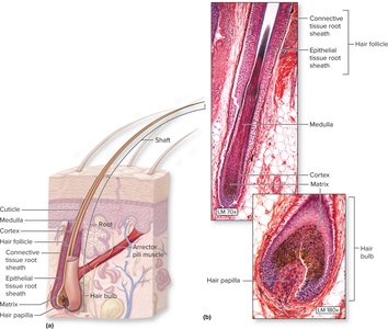
Hair is composed of dead, keratinized cells and serves protective and sensory functions:
Hair follicle: Tube-like invagination of the epidermis into the dermis.
Hair bulb: Swelling at the base of the follicle where hair production occurs.
Arrector pili muscle: Smooth muscle attached to the follicle; contraction causes "goosebumps."
Hair shaft: Portion of hair above the skin surface.
Glands of the Skin
The skin contains several types of glands with distinct functions:
Sudoriferous (sweat) glands:
Merocrine (eccrine) glands: Widely distributed; secrete watery sweat for thermoregulation.
Apocrine glands: Found in axillary and genital regions; secrete a thicker, odoriferous sweat beginning at puberty.
Sebaceous glands: Secrete sebum (oily substance) into hair follicles; lubricates and waterproofs skin and hair.
Clinical Considerations
Skin Disorders and Conditions
Acne: Inflammation of sebaceous glands and hair follicles, leading to blackheads, whiteheads, pustules, nodules, or cysts.

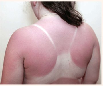
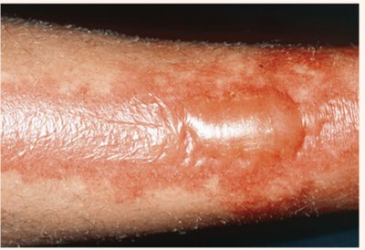
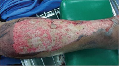
Burns: Injury to skin caused by heat, chemicals, electricity, or radiation. Classified by depth:
First-degree: Only epidermis is damaged (e.g., mild sunburn).
Second-degree: Epidermis and part of dermis are damaged; blisters may form.
Third-degree: Full thickness of skin is destroyed; may require grafting.
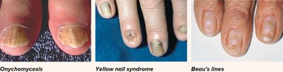
Nail disorders: Changes in nail appearance can indicate systemic disease (e.g., onychomycosis, yellow nail syndrome, Beau's lines).

Protection from UV Radiation
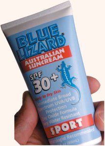
Melanin provides some protection against UV damage, but sunscreen is recommended to prevent sunburn and reduce the risk of skin cancer.
Summary Table: Layers of the Epidermis
Layer | Location | Main Features |
|---|---|---|
Stratum corneum | Surface | Dead, keratinized cells; barrier function |
Stratum lucidum | Thick skin only | Clear, dead cells; extra protection |
Stratum granulosum | Below corneum/lucidum | Keratohyalin granules; cells begin to die |
Stratum spinosum | Below granulosum | Several layers; desmosomes; Langerhans cells |
Stratum basale | Deepest | Mitotic stem cells; melanocytes; Merkel cells |
