 Back
BackThe Integumentary System: Structure and Function of the Skin
Study Guide - Smart Notes
 Tailored notes based on your materials, expanded with key definitions, examples, and context.
The Integumentary System
Overview of Skin Structure
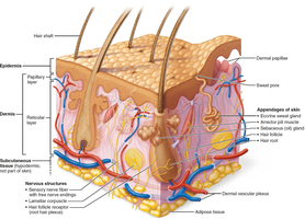
The skin, also known as the integument, is the largest organ of the human body and serves as a protective barrier. It consists of two main layers—the epidermis and the dermis—with an underlying subcutaneous layer (hypodermis) that supports skin function but is not technically part of the skin.
Epidermis: Outermost layer, composed of epithelial tissue; avascular.
Dermis: Underlies the epidermis; made of dense connective tissue; vascular.
Subcutaneous tissue (Hypodermis): Mostly adipose tissue; anchors skin, provides insulation and shock absorption.
The Epidermis
Cell Types of the Epidermis
The epidermis is primarily composed of keratinized stratified squamous epithelium and contains four main cell types, each with specialized functions:
Keratinocytes: Produce keratin, a protein that provides protective properties; most abundant cell type; continuously renewed every 25–45 days.
Melanocytes: Located in the deepest epidermis; produce melanin pigment, which protects against UV radiation.
Dendritic (Langerhans) cells: Star-shaped immune cells that patrol the epidermis and activate immune responses.
Tactile epithelial (Merkel) cells: Sensory receptors for touch, found at the epidermal-dermal junction.
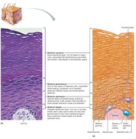
Layers of the Epidermis
The epidermis is organized into distinct layers (strata), with the number of layers varying between thick and thin skin:
Stratum basale: Deepest, single row of mitotically active stem cells; also called stratum germinativum.
Stratum spinosum: Several layers thick; contains pre-keratin filaments and desmosomes; abundant in melanosomes and dendritic cells.
Stratum granulosum: Four to six layers; cells flatten, organelles disintegrate, keratinization begins, and glycolipids are released for water resistance.
Stratum lucidum: Present only in thick skin (palms, soles); thin, translucent band of dead keratinocytes.
Stratum corneum: Outermost, 20–30 layers of dead, keratinized cells; provides protection and prevents water loss.
Thick skin contains all five layers and is found on the palms, fingertips, and soles. Thin skin lacks the stratum lucidum and is found elsewhere.
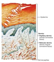
The Dermis
General Structure and Function
The dermis is a strong, flexible connective tissue layer that supports and nourishes the epidermis. It contains fibroblasts, macrophages, mast cells, and white blood cells, as well as nerves, blood vessels, lymphatics, hair follicles, and glands. The dermis is divided into two layers:
Papillary dermis: Thin, superficial layer of areolar connective tissue; contains dermal papillae with capillary loops, nerve endings, and tactile corpuscles.
Reticular dermis: Thicker, deeper layer of dense irregular connective tissue; provides strength, elasticity, and hydration; contains the dermal vascular plexus.
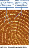
Papillary Dermis and Friction Ridges
The papillary dermis forms dermal papillae, which project into the epidermis and contain capillaries and sensory receptors. In thick skin, these papillae form friction ridges (fingerprints), which enhance grip and tactile sensitivity. The pattern of friction ridges is genetically determined and unique to each individual.
Friction ridges: Formed by dermal papillae atop dermal ridges; create epidermal ridges visible as fingerprints.
Functions: Enhance grip, amplify touch sensitivity, and produce unique fingerprint patterns via sweat pores.
Reticular Dermis
The reticular dermis constitutes the majority of the dermal thickness and is composed of dense irregular connective tissue. It contains abundant collagen and elastic fibers, which provide tensile strength, elasticity, and hydration to the skin. The dermal vascular plexus, a network of blood vessels, is found between the reticular layer and the subcutaneous tissue.
Collagen fibers: Provide structural strength and bind water for hydration.
Elastic fibers: Allow stretch and recoil of the skin.
Dermal vascular plexus: Supplies blood to the skin and supports thermoregulation.
Additional info: The reticular dermis is named for its network (reticulum) of collagen fibers, not for the presence of reticular fibers.
