 Back
BackThe Integumentary System: Structure and Function
Study Guide - Smart Notes
 Tailored notes based on your materials, expanded with key definitions, examples, and context.
The Integumentary System
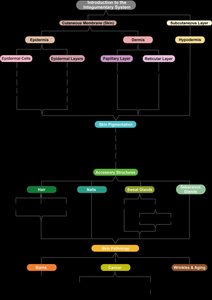
Introduction to the Integumentary System
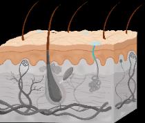
The integumentary system is an organ system composed of the skin, hair, nails, glands, and sensory receptors. It serves as the body's primary barrier to the external environment and plays a vital role in protection, sensation, and homeostasis. The skin, or cutaneous membrane, is the largest organ of the body and consists of multiple layers and accessory structures.
Main components: Epidermis, dermis, hypodermis (subcutaneous layer), hair, nails, sweat and sebaceous glands, sensory receptors.
Functions:
Protection from mechanical, chemical, UV, and microbial damage
Thermoregulation (temperature maintenance)
Sensory perception
Vitamin D synthesis
Excretion (sweat)
Expressive communication (facial expressions)
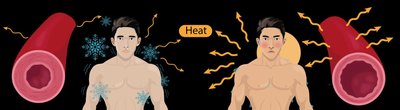
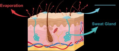
Thermoregulation in the Integumentary System
Mechanisms of Thermoregulation
The integumentary system maintains internal body temperature through two main mechanisms: vasoconstriction/vasodilation and sweating. These processes are essential for homeostasis and are regulated by negative feedback loops.
Vasoconstriction: Blood vessels constrict (decrease in diameter) when the body is cold, reducing blood flow to the skin and minimizing heat loss.
Vasodilation: Blood vessels dilate (increase in diameter) when the body is hot, increasing blood flow to the skin and facilitating heat loss.
Sweating: Sweat glands secrete a water-based solution onto the skin surface. As sweat evaporates, it cools the body.
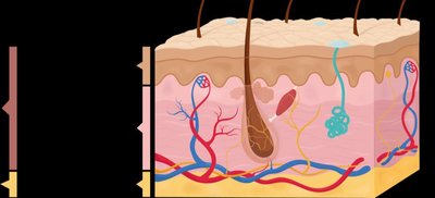
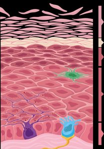
The Epidermis
Cell Types in the Epidermis
The epidermis is the outermost layer of the skin, composed of stratified squamous epithelial tissue. It contains four main cell types, each with specialized functions:
Keratinocytes: Most abundant; produce keratin, a tough, water-resistant protein that provides mechanical strength and protection.
Melanocytes: Produce melanin, a pigment that protects against UV radiation.
Dendritic (Langerhans) cells: Immune cells that help initiate immune responses and protect against pathogens.
Tactile epithelial (Merkel) cells: Work with nerve endings to detect touch sensations.
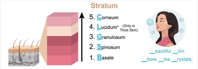
Layers of the Epidermis
The epidermis is organized into distinct layers (strata), with cells at various stages of development. The number and structure of these layers differ between thin skin (most of the body) and thick skin (palms and soles).
Stratum basale: Deepest layer; single row of dividing cells (keratinocytes, melanocytes, tactile cells).
Stratum spinosum: Several layers of keratinocytes; contains dendritic cells for immunity.
Stratum granulosum: Keratinocytes stop dividing, begin to harden and die; keratinization occurs.
Stratum lucidum: Present only in thick skin; clear, dead, densely packed cells.
Stratum corneum: Outermost layer; dead, keratin-filled cells that are regularly shed and replaced.
Thin skin: Lacks stratum lucidum, contains hair follicles and oil glands. Thick skin: Contains stratum lucidum, lacks hair follicles and oil glands, more sweat glands.
Keratinocyte Development
Keratinocytes originate in the stratum basale and are pushed upward through the layers, undergoing changes until they reach the surface and are shed. The process provides a continuous barrier and is essential for skin regeneration.
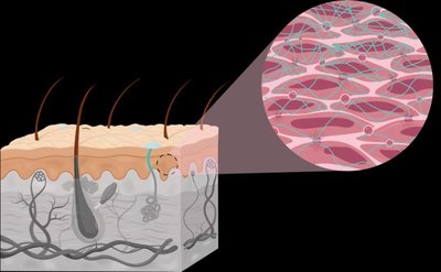
The Dermis
Structure and Layers of the Dermis
The dermis is the second, deeper layer of the skin, composed mainly of connective tissue. It provides structural support, flexibility, and houses many accessory structures.
Papillary layer: Superficial; made of loose areolar connective tissue. Contains dermal papillae (projections), capillaries, lymphatics, and Meissner corpuscles (touch receptors). Dermal papillae form epidermal ridges (fingerprints).
Reticular layer: Deep; made of dense irregular connective tissue. Contains sweat and oil glands, hair roots, Pacinian corpuscles (pressure receptors), and a network of collagen and elastic fibers. Collagen fibers form cleavage lines important for surgical incisions.

The Hypodermis (Subcutaneous Layer)
Structure and Function
The hypodermis (subcutaneous layer) lies deep to the dermis and is not technically part of the skin. It is composed mainly of adipose tissue and some areolar connective tissue. The hypodermis anchors the skin to underlying tissues, acts as a shock absorber, and insulates the body to reduce heat loss. It also serves as an energy reserve.
Functions:
Anchors skin to underlying structures
Shock absorption
Insulation and reduction of heat loss
Energy storage (fat)
Summary Table: Layers of the Skin
Layer | Main Tissue Type | Key Features | Functions |
|---|---|---|---|
Epidermis | Stratified squamous epithelium | Multiple strata, keratinocytes, melanocytes, dendritic, tactile cells | Protection, water resistance, UV protection, sensation |
Dermis (Papillary) | Areolar connective tissue | Dermal papillae, capillaries, Meissner corpuscles | Anchors epidermis, sensation, fingerprints |
Dermis (Reticular) | Dense irregular connective tissue | Collagen/elastic fibers, glands, hair roots, Pacinian corpuscles | Strength, flexibility, houses accessory structures |
Hypodermis | Adipose & areolar tissue | Fat storage, loosely anchors skin | Insulation, shock absorption, energy reserve |
