 Back
BackThe Integumentary System: Structure and Function
Study Guide - Smart Notes
 Tailored notes based on your materials, expanded with key definitions, examples, and context.
The Integumentary System
Overview of the Integumentary System
The integumentary system is composed of the skin and its accessory structures, including hair, nails, and glands. It serves as the body's primary barrier against the external environment and plays critical roles in protection, sensation, thermoregulation, and synthesis of vitamin D.
Skin: The largest organ of the body, consisting of multiple layers with specialized cells and structures.
Accessory Structures: Include hair, nails, sweat glands, and sebaceous (oil) glands.
Main Functions: Protection, temperature regulation, sensory reception, excretion, and metabolic functions.
Layers of the Skin
Epidermis, Dermis, and Hypodermis
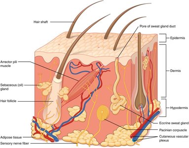
The skin is organized into three main layers: the epidermis, dermis, and hypodermis. Each layer has distinct structures and functions.
Epidermis: The outermost layer, composed mainly of keratinized stratified squamous epithelium. It provides a waterproof barrier and creates our skin tone.
Dermis: The middle layer, containing connective tissue, blood vessels, nerve endings, hair follicles, and glands. It supports and nourishes the epidermis.
Hypodermis (Subcutaneous Layer): The deepest layer, consisting mainly of adipose tissue. It insulates the body and anchors the skin to underlying structures.
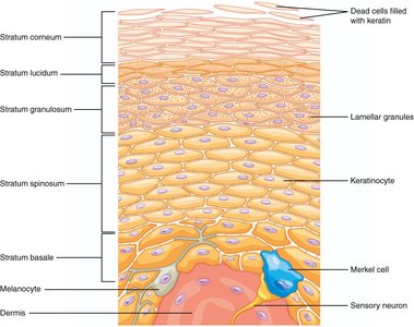
Microscopic Structure of the Epidermis
Layers of the Epidermis
The epidermis is composed of several distinct layers (strata), each with specialized cells and functions. From deep to superficial, these layers are:
Stratum Basale: The deepest layer, containing stem cells that divide to form new keratinocytes. Also contains melanocytes (produce pigment) and Merkel cells (sensory receptors).
Stratum Spinosum: Several layers of keratinocytes connected by desmosomes; provides strength and flexibility.
Stratum Granulosum: Cells begin to flatten and accumulate keratohyalin granules, which contribute to waterproofing.
Stratum Lucidum: Present only in thick skin (palms, soles); a thin, clear layer of dead keratinocytes.
Stratum Corneum: The outermost layer, consisting of dead, flattened, keratin-filled cells that are continuously shed and replaced.
Accessory Structures of the Skin
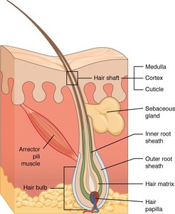
Hair and Hair Follicles
Hair is a filamentous structure composed of keratinized cells. It arises from hair follicles, which are complex structures extending from the epidermis into the dermis.
Hair Shaft: The visible part of the hair above the skin surface.
Hair Root: The portion of hair below the skin surface, within the follicle.
Hair Bulb: The expanded base of the hair follicle, containing the hair matrix (site of cell division) and hair papilla (provides nutrients).
Medulla, Cortex, Cuticle: Layers of the hair shaft; the medulla is the central core, the cortex surrounds the medulla, and the cuticle is the outermost layer.
Arrector Pili Muscle: A small muscle attached to the hair follicle; contraction causes "goosebumps."
Sebaceous Gland: Produces sebum (oil) that lubricates hair and skin.
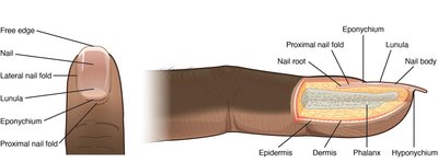
Nails
Nails are protective coverings on the dorsal surface of the distal fingers and toes. They are composed of hard, keratinized cells.
Nail Body: The visible, attached part of the nail.
Lunula: The whitish, crescent-shaped area at the base of the nail.
Eponychium (Cuticle): The fold of skin that overlaps the nail at the base.
Hyponychium: The area under the free edge of the nail, providing a barrier to infection.
Nail Root: The portion of the nail embedded under the skin, where nail growth occurs.
Glands of the Skin
Types of Skin Glands
The skin contains several types of glands, each with specific functions:
Eccrine (Merocrine) Sweat Glands: Widely distributed; secrete watery sweat for thermoregulation.
Apocrine Sweat Glands: Located mainly in axillary and genital areas; secrete a thicker fluid into hair follicles, active after puberty.
Sebaceous (Oil) Glands: Associated with hair follicles; secrete sebum to lubricate and waterproof skin and hair.
Example: Eccrine sweat glands are essential for regulating body temperature through evaporative cooling during exercise or exposure to heat.
Summary Table: Layers and Structures of the Skin
Layer/Structure | Main Components | Function |
|---|---|---|
Epidermis | Keratinocytes, melanocytes, Merkel cells | Protection, waterproofing, sensation |
Dermis | Connective tissue, blood vessels, nerves, glands, hair follicles | Support, nourishment, sensation, thermoregulation |
Hypodermis | Adipose tissue | Insulation, energy storage, anchoring skin |
Hair Follicle | Hair bulb, matrix, papilla, arrector pili muscle | Hair production, sensation, minor thermoregulation |
Nail | Nail body, lunula, eponychium, hyponychium | Protection, fine manipulation |
Sweat Glands | Eccrine, apocrine glands | Thermoregulation, excretion |
Sebaceous Glands | Sebum (oil) | Lubrication, waterproofing |
