 Back
BackThe Integumentary System: Structure and Function
Study Guide - Smart Notes
 Tailored notes based on your materials, expanded with key definitions, examples, and context.
The Integumentary System
Overview and Components
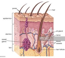
The integumentary system is the body's largest organ system, consisting of the skin and its accessory structures such as hair, glands, and nails. It serves as a protective barrier, regulates temperature, and provides sensory information.
Skin: Composed of multiple tissue types, including epithelial, connective, muscle, and nervous tissue.
Accessory Structures: Hair, glands (sebaceous and sweat), and nails.
Skin Layers
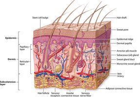
The skin is composed of two main layers: the epidermis and the dermis. Beneath the dermis lies the hypodermis (subcutaneous tissue), which connects the skin to underlying tissues but is not considered part of the integument.
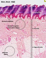
Epidermis: Outermost layer, composed of stratified squamous keratinized epithelium.
Dermis: Deeper layer, composed of connective tissue, blood vessels, and sensory receptors.
Hypodermis: Subcutaneous layer containing adipose tissue for insulation and energy storage.
The Epidermis
Layers of the Epidermis
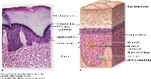
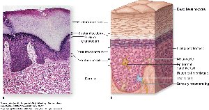
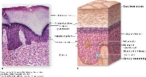
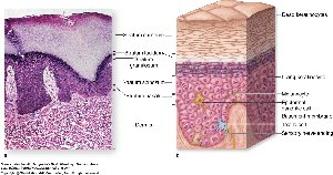
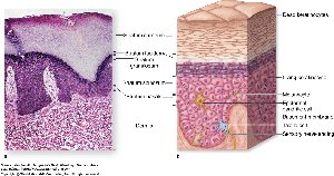
The epidermis consists of several layers (strata) of cells, each with distinct functions. From superficial to deep, the layers are:
Stratum Corneum: Superficial layer of dead, keratinized cells (keratinocytes) that provide a tough, protective barrier.
Stratum Lucidum: Thin, translucent layer of dead cells found only in thick skin (palms and soles).
Stratum Granulosum: Contains keratinocytes with granules that release waterproofing substances.
Stratum Spinosum: First layer with metabolically active cells; contains melanin for UV protection.
Stratum Basale: Deepest layer; single row of dividing cells, including melanocytes and Merkel cells.
Specialized Cells of the Epidermis
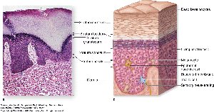
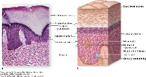
Keratinocytes: Main cell type, produce keratin for protection.
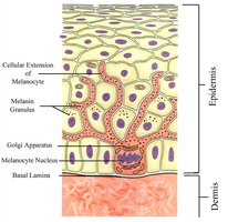
Melanocytes: Produce melanin pigment, found in the stratum basale.
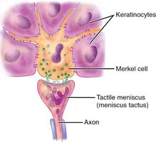
Merkel Cells: Sensory cells for light touch, also in the stratum basale.
Basement Membrane
The basement membrane is located immediately deep to the stratum basale. It consists of the basal lamina (produced by keratinocytes) and the reticular lamina (produced by the dermis). This structure anchors the epidermis to the dermis and prevents separation.
Mnemonic for Epidermal Layers
To remember the order of the epidermal layers (superficial to deep): Come, Let's Get Sun Burned (Corneum, Lucidum, Granulosum, Spinosum, Basale).
Cell Death in the Epidermis
The epidermis is avascular, so only cells near the dermis (stratum basale, spinosum, and lower granulosum) receive nutrients and oxygen. As cells migrate outward, they die and become keratinized.
The Dermis
Layers of the Dermis
The dermis is a connective tissue layer beneath the epidermis, divided into two regions:
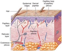
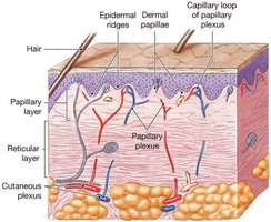
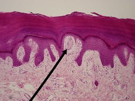
Papillary Layer: Superficial, composed of loose connective tissue. Contains dermal papillae (fingerlike projections) that form fingerprints and house tactile (Meissner's) corpuscles and capillary loops.
Reticular Layer: Deeper, thicker layer of dense irregular connective tissue. Contains sweat glands, sebaceous glands, blood vessels, lamellated (Pacinian) corpuscles, and free nerve endings for pain sensation.
Accessory Structures of the Skin
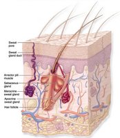
Hair
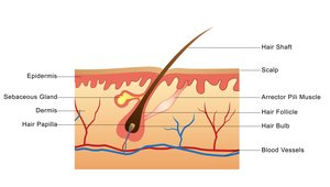
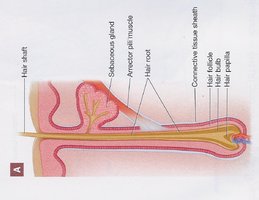
Hair consists of dead, keratinized cells and is divided into:
Shaft: Visible part above the skin surface.
Root: Embedded in the dermis, surrounded by the hair follicle.
Arrector Pili Muscle: Smooth muscle that causes hair to stand erect when contracted (goosebumps).
Nails
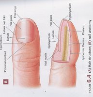
Nails are protective coverings composed of dead, keratinized cells. Main parts include:
Nail Plate: Main body of the nail.
Nail Folds: Skin folds on three sides of the nail.
Nail Matrix: Proximal region where nail growth occurs.
Lunula: Visible white crescent at the nail base.
Eponychium: Cuticle, skin covering the nail plate.
Hyponychium: Skin under the free edge of the nail.
Glands
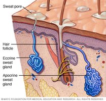
Sebaceous Glands: Exocrine glands that secrete sebum (oil) into hair follicles for lubrication.
Sweat Glands:
Apocrine: Found in armpits and groin; secrete fatty sweat that bacteria break down, causing odor.
Eccrine (Merocrine): Most common; found all over the body and secrete watery sweat for thermoregulation.
Thin Skin vs. Thick Skin
Comparison of Thin and Thick Skin
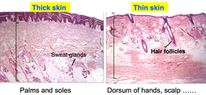
Feature | Thick Skin | Thin Skin |
|---|---|---|
Location | Palms, soles | Everywhere else |
Layers | 5 (includes stratum lucidum) | 4 (no stratum lucidum) |
Hair/Sebaceous Glands | Absent | Present |
Dermis Thickness | Thinner | Thicker |
Dermal Papillae | More pronounced | Less pronounced |
Sensory Receptors in the Skin
Types of Sensory Receptors
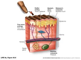
Tactile (Meissner's) Corpuscles: Detect fine touch, located in dermal papillae near the skin surface.
Merkel Disks: Specialized cells in the stratum basale for light touch, especially in fingertips.
Pacinian (Lamellated) Corpuscles: Detect deep pressure and vibration, located in the reticular dermis and hypodermis.
Free Nerve Endings: Detect pain, temperature, and crude touch.
Summary Table: Structures to Identify in the Skin
Structure | Location/Function |
|---|---|
Epidermis | Outer protective layer |
Dermis | Connective tissue, supports epidermis |
Stratum Corneum | Dead, keratinized cells |
Stratum Lucidum | Only in thick skin |
Stratum Granulosum | Keratin granules, waterproofing |
Stratum Spinosum | Melanin, active cells |
Stratum Basale | Dividing cells, melanocytes, Merkel cells |
Papillary Layer | Dermal papillae, Meissner's corpuscles |
Reticular Layer | Sweat/sebaceous glands, Pacinian corpuscles |
Arrector Pili | Hair erection |
Hair Follicle/Bulb/Root/Shaft | Hair growth and structure |
Eccrine Sweat Gland | Thermoregulation |
Apocrine Sweat Gland | Odorous sweat |
Sebaceous Gland | Secretes sebum |
Meissner's Corpuscle | Fine touch |
Pacinian Corpuscle | Pressure/vibration |
