 Back
BackThe Integumentary System: Structure, Function, and Clinical Relevance
Study Guide - Smart Notes
 Tailored notes based on your materials, expanded with key definitions, examples, and context.
The Integumentary System
Overview and Learning Outcomes
The integumentary system, primarily composed of the skin and its appendages, is the largest organ system of the human body. It serves as a protective barrier, regulates body temperature, and provides sensory information. Mastery of anatomical vocabulary, clinical reasoning, and the ability to connect structure to function are essential for students pursuing health-related careers.
Build Foundational Anatomical Vocabulary & Concepts: Accurate use of anatomical and clinical terms is essential for communication in health sciences.
Connect Anatomy and Physiology to Health Careers: Understanding the integumentary system is crucial for clinical practice and patient care.
Develop Clinical Reasoning: Scenario analysis and case studies help predict and justify clinical findings.
Foster Peer Collaboration: Teamwork and discussion enhance understanding of scientific concepts.
Support Professional Exam Readiness: Objectives align with board-style and licensure exam requirements.
Skin Structure and Layers
Gross Structure of the Skin
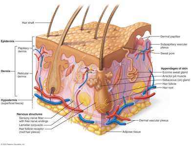
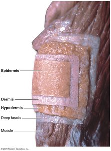
The skin consists of three primary layers: the epidermis, dermis, and hypodermis (subcutaneous tissue). Each layer has distinct structural and functional properties.
Epidermis: The outermost, avascular layer composed mainly of keratinized stratified squamous epithelium.
Dermis: A connective tissue layer beneath the epidermis, containing blood vessels, nerves, and appendages.
Hypodermis (Superficial Fascia): A layer of adipose and areolar tissue that anchors skin to underlying structures and provides insulation.
Functions of the Skin
Protection: Acts as a barrier against mechanical injury, pathogens, and harmful substances.
Thermoregulation: Regulates body temperature through sweat production and blood flow.
Sensation: Contains sensory receptors for touch, pain, temperature, and pressure.
Metabolic Functions: Synthesizes vitamin D when exposed to UV light.
Excretion: Eliminates waste products through sweat.
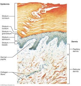
Microscopic Structure of the Epidermis
Layers of the Epidermis
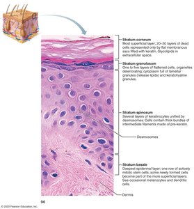
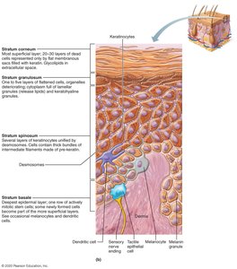
The epidermis is composed of several distinct layers, each with specialized cells and functions. From superficial to deep, these layers are:
Stratum Corneum: 20–30 layers of dead, keratinized cells that provide a tough, protective outer layer.
Stratum Granulosum: Cells contain keratohyalin granules and begin to lose their nuclei as they move upward.
Stratum Spinosum: Several layers of keratinocytes unified by desmosomes; cells contain thick bundles of intermediate filaments.
Stratum Basale: Deepest layer; a single row of actively mitotic stem cells, melanocytes, and some dendritic cells.
Additional info: In thick skin (palms, soles), a fifth layer called the stratum lucidum is present between the stratum corneum and stratum granulosum.
Dermis and Its Modifications
Structure of the Dermis
The dermis is divided into two layers:
Papillary Dermis: Thin, superficial layer composed of areolar connective tissue; forms dermal papillae that interdigitate with the epidermis.
Reticular Dermis: Thicker, deeper layer composed of dense irregular connective tissue; contains collagen and elastic fibers for strength and flexibility.
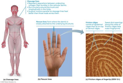
Dermal Modifications:
Cleavage (Tension) Lines: Indicate the orientation of collagen fibers; important for surgical incisions.
Flexure Lines: Occur where the dermis is tightly attached to underlying structures, such as palms and fingers.
Friction Ridges: Form fingerprints; enhance grip and tactile sensation.
Appendages of the Skin
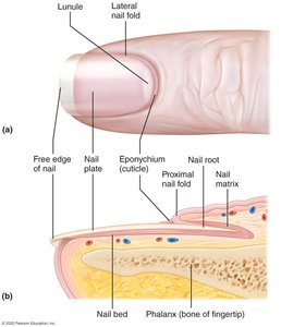
Nails
Nails are hard, keratinized structures that protect the distal tips of fingers and toes and aid in manipulation of objects.
Nail Plate: Visible, hard part of the nail.
Nail Matrix: Proximal region responsible for nail growth.
Eponychium (Cuticle): Fold of skin at the base of the nail.
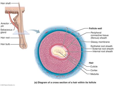
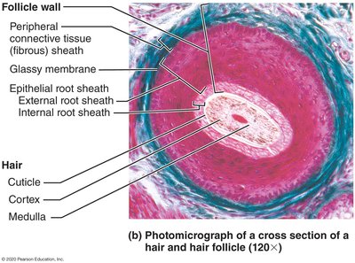
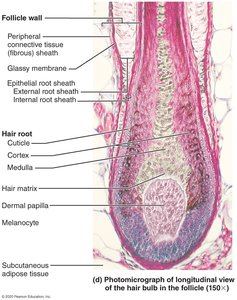
Hair and Hair Follicles
Hair is composed of dead, keratinized cells and serves protective and sensory functions. Hair follicles are complex structures that anchor each hair into the skin.
Hair Shaft: Visible part above the skin surface.
Hair Root: Portion within the follicle below the skin surface.
Hair Bulb: Expanded base of the follicle where hair growth occurs.
Associated Structures: Arrector pili muscle (causes hair to stand), sebaceous glands (secrete sebum).
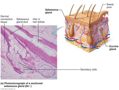
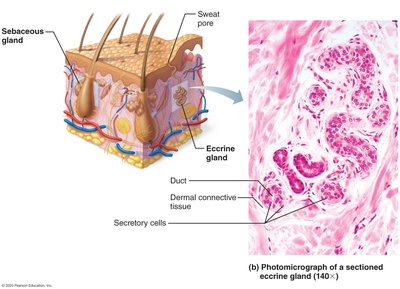
Skin Glands
The skin contains several types of glands with distinct functions:
Sebaceous (Oil) Glands: Secrete sebum into hair follicles to lubricate skin and hair.
Eccrine (Sweat) Glands: Widely distributed; produce watery sweat for thermoregulation.
Apocrine Glands: Located in axillary and genital areas; secrete a thicker fluid into hair follicles, active after puberty.
Clinical Correlations
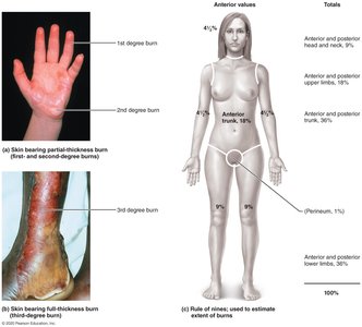
Burns
Burns are classified by depth and extent of tissue damage. The "rule of nines" is used to estimate the percentage of body surface area affected.
First-Degree Burns: Affect only the epidermis; cause redness and pain.
Second-Degree Burns: Involve the epidermis and part of the dermis; cause blistering.
Third-Degree Burns: Destroy the entire thickness of the skin; require medical intervention.
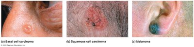
Skin Cancers
Skin cancers are abnormal growths of skin cells, often caused by UV exposure. The three main types are:
Basal Cell Carcinoma: Most common, least malignant; arises from basal cells.
Squamous Cell Carcinoma: Arises from keratinocytes of the stratum spinosum; can metastasize if untreated.
Melanoma: Most dangerous; arises from melanocytes and is highly metastatic.
ABCDE Rule for Melanoma Detection:
Asymmetry
Border irregularity
Color variation
Diameter > 6 mm
Evolving shape or color
Summary Table: Layers of the Skin
Layer | Main Components | Functions |
|---|---|---|
Epidermis | Keratinocytes, melanocytes, dendritic cells | Protection, waterproofing, UV defense |
Dermis | Collagen, elastic fibers, blood vessels, nerves | Support, sensation, thermoregulation |
Hypodermis | Adipose tissue, areolar tissue | Insulation, energy storage, anchoring skin |
Key Terms and Concepts
Keratin: A tough, fibrous protein that provides waterproofing and protection.
Melanin: Pigment produced by melanocytes; protects against UV radiation.
Desmosomes: Cell junctions that hold keratinocytes together.
Dermal Papillae: Projections that increase surface area for exchange of gases, nutrients, and waste products between layers.
