 Back
BackThe Integumentary System: Structure, Function, and Clinical Aspects
Study Guide - Smart Notes
 Tailored notes based on your materials, expanded with key definitions, examples, and context.
The Integumentary System
Overview and Major Components
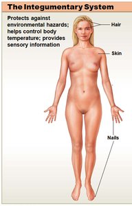
The integumentary system is the body's largest organ system, serving as the primary barrier between the internal environment and the external world. It consists of the cutaneous membrane (skin) and accessory structures such as hair, nails, and exocrine glands.
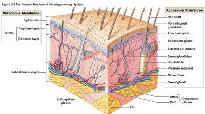
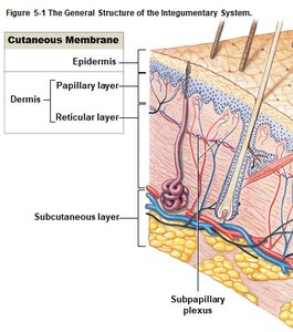
Cutaneous membrane (skin): Composed of the epidermis and dermis.
Accessory structures: Include hair, nails, and exocrine glands (sebaceous and sweat glands).
Subcutaneous Layer (Hypodermis)
The subcutaneous layer, or hypodermis, is a loose connective tissue layer beneath the dermis. It is not technically part of the integumentary system but is closely associated with it.
Separates the skin from deeper tissues and organs.
Contains adipose tissue for insulation and energy storage.
Common site for subcutaneous injections due to absence of vital organs.
Functions of the Integumentary System
General Functions
Protection: Shields underlying tissues from mechanical, chemical, and biological damage; prevents fluid loss.
Temperature maintenance: Regulates heat exchange with the environment.
Synthesis and storage of nutrients: Epidermis synthesizes vitamin D3; dermis stores lipids.
Sensory reception: Contains receptors for touch, pressure, pain, and temperature.
Excretion and secretion: Glands excrete salts, water, and organic wastes; mammary glands secrete milk.
The Epidermis
Structure and Cell Types
The epidermis is a stratified epithelial tissue that is avascular and relies on diffusion from the underlying dermis for nutrients. The majority of cells are keratinocytes, which produce the protein keratin.
Keratinocytes: Main cell type, produce keratin for waterproofing and protection.
Melanocytes: Produce melanin pigment for UV protection.
Tactile (Merkel) cells: Sensory receptors for touch.
Thick Skin vs. Thin Skin
Thick skin: Found on palms and soles; has five layers, including a prominent stratum corneum.
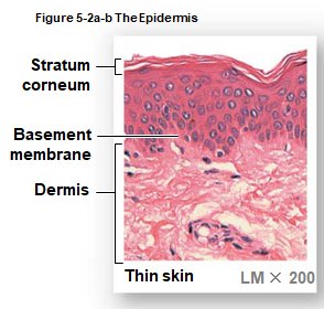
Thin skin: Covers most of the body; has four layers and a thinner stratum corneum.
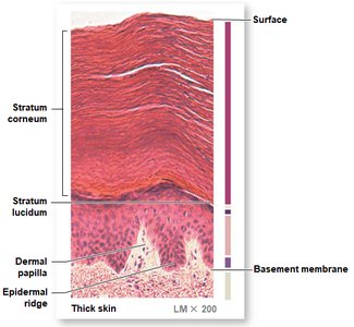
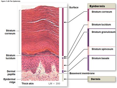
Strata of the Epidermis
The epidermis is organized into distinct layers (strata), each with specialized functions:
Stratum basale (germinativum): Deepest layer; contains stem cells, melanocytes, and tactile cells. Site of cell division.
Stratum spinosum: Several layers of keratinocytes; contains dendritic cells for immune defense.
Stratum granulosum: Cells stop dividing and begin producing keratin.
Stratum lucidum: Present only in thick skin; clear, densely packed cells filled with keratin.
Stratum corneum: Outermost layer; 15–30 layers of dead, keratinized cells that are shed in sheets.
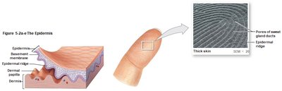
Epidermal Ridges and Dermal Papillae
Epidermal ridges (from the stratum basale) interlock with dermal papillae, forming the basis for fingerprints and enhancing grip.
Skin Color and Pigmentation
Pigments
Carotene: Orange-yellow pigment from diet, accumulates in epidermal cells.
Melanin: Brown-black or red-yellow pigment produced by melanocytes; stored in melanosomes and transferred to keratinocytes.
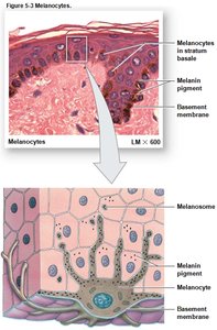
Role of Sunlight
UV exposure increases melanin production, protecting deeper tissues.
Sunlight enables synthesis of vitamin D3, which is converted to calcitriol for calcium and phosphorus absorption.
Dermal Circulation and Skin Color
Oxygenated blood gives skin a reddish tint; vasodilation causes flushing, vasoconstriction causes paleness.
Cyanosis (bluish color) indicates low oxygen levels.
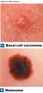
Skin Cancer
Basal cell carcinoma: Most common, originates in stratum basale.
Squamous cell carcinoma: Arises from superficial layers.
Malignant melanoma: Most dangerous, can metastasize rapidly.
The Dermis
Layers of the Dermis
Papillary layer: Superficial, areolar tissue; contains capillaries, lymphatics, and sensory neurons.
Reticular layer: Deeper, dense irregular connective tissue; contains collagen and elastic fibers, blood vessels, nerves, and accessory organs.
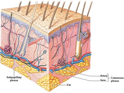
Blood Supply
Cutaneous plexus: Supplies blood to the hypodermis and deeper dermis.
Subpapillary plexus: Supplies blood to the superficial dermis and epidermis.
Accessory Structures
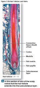
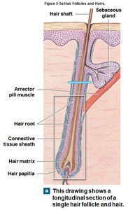
Hair and Hair Follicles
Hair is a nonliving, keratinized structure produced by hair follicles. It serves protective, sensory, and thermoregulatory functions.
Hair follicle: Extends into the dermis and hypodermis; contains hair matrix (site of cell division) and hair papilla (connective tissue with capillaries and nerves).
Hair structure: Root (below skin), shaft (visible part), cuticle (outer layer), cortex (middle), medulla (core).
Hair Growth and Color
Hair grows in cycles; growth phase followed by rest phase.
Color determined by type and amount of melanin; graying occurs with age as pigment production declines.
Exocrine Glands of the Skin
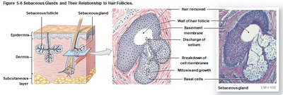
Sebaceous (oil) glands: Secrete sebum into hair follicles or directly onto skin; lubricates and protects.
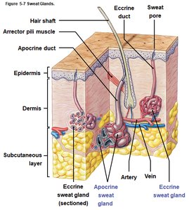
Apocrine sweat glands: Found in armpits, nipples, and pubic region; produce odorous secretion.
Eccrine (merocrine) sweat glands: Widely distributed; secrete watery sweat for thermoregulation.
Modified sweat glands: Mammary glands (milk), ceruminous glands (earwax).
Nails
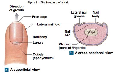
Nails are protective structures on the dorsal surfaces of fingers and toes, composed of dense, keratinized cells.
Nail body: Visible part, covers the nail bed.
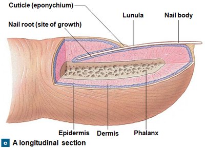
Nail root: Site of nail production, covered by the cuticle (eponychium).
Lunula: Pale crescent near the root.
Repair and Regeneration of the Integument
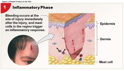
Phases of Skin Repair
Inflammatory phase: Bleeding and inflammation occur; mast cells trigger response.
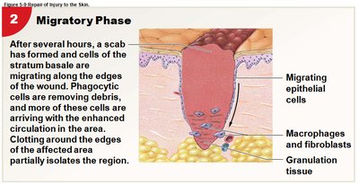
Migratory phase: Scab forms; epithelial cells migrate; phagocytes remove debris; granulation tissue forms.
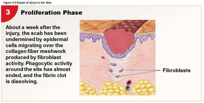
Proliferation phase: Epidermal cells migrate over collagen meshwork; fibroblasts produce new tissue.
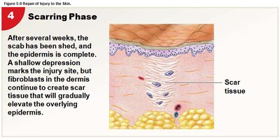
Scarring phase: Scab is shed; scar tissue forms, elevating the epidermis.
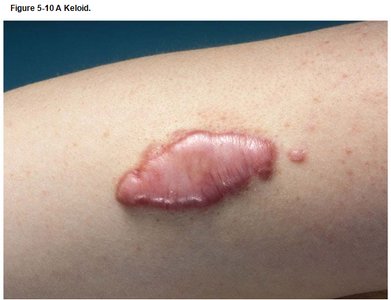
Keloids
Keloids are thickened areas of scar tissue with a shiny, smooth surface, resulting from excessive collagen formation during healing.
Clinical Aspects: Burns and Aging
Burns
First-degree (superficial) burns: Affect only the epidermis; cause redness and mild pain (e.g., sunburn).
Second-degree (partial-thickness) burns: Damage epidermis and part of dermis; cause blistering and pain.
Third-degree (full-thickness) burns: Destroy epidermis and dermis; may extend into hypodermis; require skin grafting.
Burns compromise fluid balance, thermoregulation, and protection from infection.
Effects of Aging
Epidermis thins; stem cell activity declines.
Immune response weakens due to fewer macrophages.
Vitamin D3 production decreases, affecting bone strength.
Melanin production declines, increasing sun sensitivity.
Glandular activity decreases, causing dry, scaly skin.
Hair thins and changes color; skin becomes less elastic and more prone to injury.
Skin repair slows, increasing infection risk.
