 Back
BackThe Integumentary System: Structure, Function, and Components
Study Guide - Smart Notes
 Tailored notes based on your materials, expanded with key definitions, examples, and context.
Module 5.1 Overview of the Integumentary System
Skin Structure
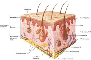
The skin is the largest organ of the body, accounting for 10–15% of total body weight. It serves as more than just an outer covering, functioning as a complex organ system with multiple layers and accessory structures.
Cutaneous membrane (skin): Composed of two main layers:
Epidermis: The superficial layer, consisting of keratinized stratified squamous epithelium resting on a basement membrane. It is avascular and relies on diffusion from the dermis for nutrients and oxygen.
Dermis: Located deep to the epidermis and basement membrane, made of loose connective tissue and dense irregular connective tissue. It is highly vascular and contains sensory receptors.
Accessory structures: Embedded in the cutaneous membrane, including sweat glands, sebaceous glands, hair, and nails.
Hypodermis (superficial fascia or subcutaneous fat): Not part of the skin, but anchors it to deeper structures like muscle and bone. Composed of loose connective and adipose tissues with abundant blood supply.
Functions of the Integumentary System
The integumentary system is critical for protecting underlying organs and maintaining homeostasis through several key functions:
Protection: Provides a barrier against mechanical trauma, pathogens, and environmental hazards (e.g., UV light). The acid mantle (slightly acidic pH from sebaceous gland secretions) inhibits pathogen growth. Hydrophobic lipid-based chemicals repel water and salts, maintaining water and electrolyte balance.
Sensation: Contains numerous sensory receptors that detect changes in the internal and external environment, including heat, cold, and pain.
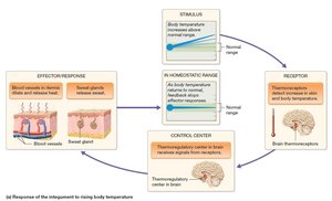
Thermoregulation: Maintains stable internal temperature via negative feedback loops. The hypothalamus acts as the thermoregulatory center, initiating sweating and vasodilation in response to heat, and vasoconstriction in response to cold.
Excretion: Eliminates waste products and toxins from the body through sweat.
Vitamin D Synthesis: The skin is involved in the synthesis of vitamin D when exposed to UV light.
Module 5.2 The Epidermis
The Epidermis: Cell Types and Layers
The epidermis is the most superficial layer of the skin and is composed of several specialized cell types and distinct layers.
Keratinocytes: Make up about 95% of the epidermis. They manufacture keratin, a tough fibrous protein, and are linked by desmosomes.
Dendritic (Langerhans) cells: Located in the stratum spinosum; act as phagocytes of the immune system.
Merkel cells: Found in the stratum basale; sensory receptors for light touch, especially numerous in fingertips, lips, and hair bases.
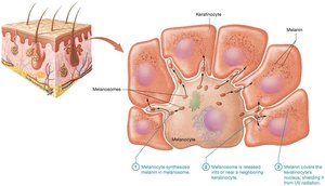
Melanocytes: Located in the stratum basale; produce melanin, a pigment that protects against UV radiation.
Layers of the Epidermis (from deep to superficial):
Stratum basale (germinativum): Single layer of stem cells; most metabolically active; responsible for vitamin D synthesis and replacement of dead keratinocytes.
Stratum spinosum: Thickest layer; metabolically and mitotically active.
Stratum granulosum: 3–5 layers; contains keratin bundles and lipid-based granules for waterproofing.
Stratum lucidum: Narrow layer of clear, dead keratinocytes; present only in thick skin.
Stratum corneum: Outermost layer; several layers of dead, flattened keratinocytes filled with keratin; cells are sloughed off mechanically.
Mnemonic: "Brilliant Studying Gives Loads of Confidence" (Basale, Spinosum, Granulosum, Lucidum, Corneum)
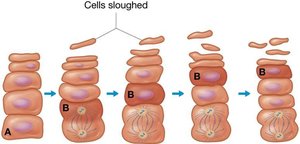
Keratinocyte Life Cycle
Keratinocytes are continuously produced in the stratum basale and spinosum, migrate upward, and are eventually shed from the stratum corneum. This process takes 40–50 days.
Module 5.3 The Dermis
The Dermis: Structure and Function
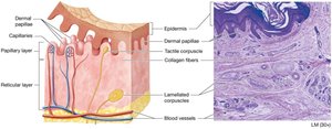
The dermis is a highly vascular layer beneath the epidermis, providing structural support, nourishment, and sensory functions. It consists of two distinct layers:
Papillary layer: Thinner, superficial layer made of loose connective tissue. Contains dermal papillae, which anchor the epidermis and contain capillaries and tactile (Meissner) corpuscles for light touch sensation.
Reticular layer: Deeper, thicker layer composed of dense irregular connective tissue. Contains collagen bundles for strength, elastic fibers for flexibility, and proteoglycans for hydration. Also houses lamellated (Pacinian) corpuscles (pressure/vibration receptors), blood vessels, sweat glands, hair, sebaceous glands, and adipose tissue.

Skin Markings and Wrinkles
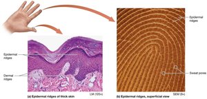
Dermal ridges: Prominent areas of dermal papillae that create epidermal ridges (fingerprints), enhancing grip. Patterns are genetically determined and unique.
Skin wrinkles: Result from decreased collagen, elastic fibers, proteoglycans, and adipose tissue with age. Accelerated by UV exposure and smoking. Treatments include Botox, fillers, and topical creams (with limited effectiveness).
Module 5.4 Skin Pigmentation
Melanin and Skin Color
Melanin is produced by melanocytes in the stratum basale and is the primary determinant of skin color. It protects keratinocyte DNA from UV-induced mutations and must be continuously produced.
Synthesis: Melanin is synthesized from tyrosine via the enzyme tyrosinase within melanosomes.
UV exposure: Increases melanin production, leading to tanning. Both UVA and UVB rays can damage DNA and increase cancer risk.
Vitamin D regulation: Melanin reduces vitamin D synthesis, affecting calcium absorption. Populations in high UV regions evolved darker skin to prevent excess vitamin D; those in low UV regions evolved lighter skin for adequate synthesis.
Distribution: All humans have similar numbers of melanocytes; differences in skin color are due to tyrosinase activity and melanin type.
Common Pigmentation Variations
Freckle: Localized increased melanin production.
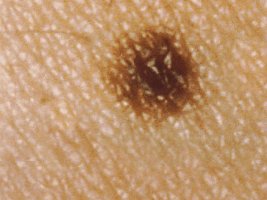
Mole (nevus): Local proliferation of melanocytes.
Albinism: Genetic lack of tyrosinase, resulting in little or no melanin and increased UV sensitivity.

Other Pigments Affecting Skin Color
Carotene: Yellow-orange pigment from diet (e.g., egg yolks, orange vegetables).
Hemoglobin: Red pigment in blood; visible through the skin, especially with increased blood flow (erythema) or decreased oxygenation (cyanosis).
Module 5.5 Accessory Structures of the Integument: Hair, Nails, and Glands
Hair: Structure and Function
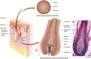
Hair is a filamentous structure found on most body surfaces, providing protection and sensory input. It is composed of keratinized epithelial cells in various stages of development.
Shaft: Portion above the skin surface; made of dead, keratinized cells.
Root: Embedded in the dermis; contains living cells and is surrounded by the hair follicle. The base (hair bulb) contains the hair papilla (blood supply).
Layers of hair (transverse section):
Medulla: Soft core (only in thick hair).
Cortex: Several layers of keratinocytes with hard keratin.
Cuticle: Outermost layer of overlapping keratinocytes.
Arrector pili muscle: Smooth muscle that causes hair to stand up (goosebumps).
Hair Types and Pigmentation
Lanugo: Thin, nonpigmented fetal hair.
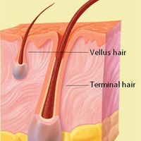
Terminal hair: Thick, coarse, pigmented (scalp, eyebrows, eyelashes, after puberty in certain regions).
Vellus hair: Thin, nonpigmented, covers most of the body.
Hair color: Determined by melanin type and amount; less melanin with age leads to gray/white hair.
Glands of the Skin
Sudoriferous (sweat) glands: Two main types:
Eccrine glands: Widely distributed; secrete watery sweat for thermoregulation.
Apocrine glands: Found in axillae, anal region, and areolae; secrete thick, protein-rich sweat into hair follicles; odor develops from bacterial action; secretion influenced by sex hormones.
Sebaceous glands: Secrete oily sebum onto hair follicles; use holocrine secretion; provide a hydrophobic barrier and deter bacteria; most abundant on face and scalp.
Ceruminous glands: Modified apocrine glands in the ear; produce cerumen (ear wax) to trap particles and lubricate the ear canal.
Mammary glands: Specialized sweat glands that produce milk.

Gland Type | Location | Secretion | Function |
|---|---|---|---|
Eccrine sweat gland | Throughout skin | Watery sweat (99% water + electrolytes) | Thermoregulation |
Apocrine sweat gland | Axillae, anal region, areolae | Thick, protein-rich sweat | Odor (bacterial action), pheromonal communication |
Sebaceous gland | Face, scalp, associated with hair follicles | Sebum (oil) | Hydrophobic barrier, antibacterial |
Ceruminous gland | Ear canal | Cerumen (ear wax) | Traps particles, lubricates |
Mammary gland | Breast tissue | Milk | Nourishment for infants |
