 Back
BackThe Integumentary System: Structure, Function, and Clinical Relevance
Study Guide - Smart Notes
 Tailored notes based on your materials, expanded with key definitions, examples, and context.
Overview of the Integumentary System
Introduction
The integumentary system is composed of the skin and its accessory structures, including hair, nails, and glands. It serves as the body's primary barrier against the external environment and plays critical roles in protection, sensation, thermoregulation, excretion, and vitamin D synthesis.

Skin Structure—Basic Anatomy
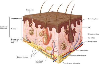
Layers of the Skin
Epidermis: The outermost layer, composed of keratinized stratified squamous epithelium. It provides a waterproof barrier and creates our skin tone.
Dermis: The middle layer, consisting of connective tissue, blood vessels, nerve endings, hair follicles, and glands. It provides structural strength and elasticity.
Hypodermis (subcutaneous layer): Not technically part of the skin, this layer consists mainly of adipose tissue and anchors the skin to underlying tissues.
Functions of the Integumentary System
Protection: Acts as a barrier against mechanical trauma, pathogens, and environmental hazards. Keratin and melanin provide toughness and UV protection.
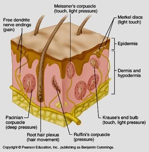
Sensation: Contains sensory receptors for pain, temperature, touch, and pressure.
Thermoregulation: Regulates body temperature through blood flow and sweat production.
Excretion: Eliminates small amounts of waste products via sweat.
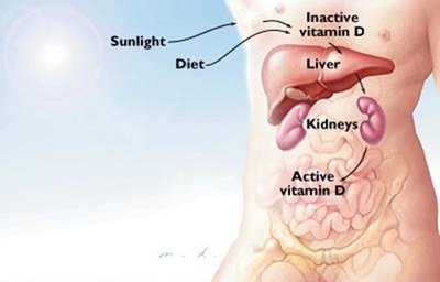
Vitamin D Synthesis: Initiates the production of vitamin D when exposed to UV light.
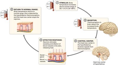
Thermoregulation
Mechanisms of Temperature Regulation
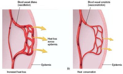
Vasodilation: Blood vessels in the dermis widen, increasing blood flow to the skin and promoting heat loss.
Vasoconstriction: Blood vessels narrow, reducing blood flow to the skin and conserving heat.
Sweating: Sweat glands produce sweat, which evaporates and cools the body.
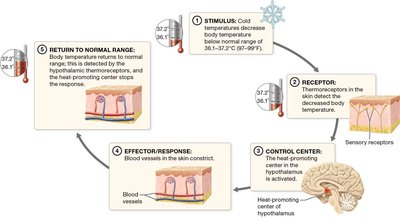
Thermoregulatory Feedback Loops
Negative feedback mechanisms maintain body temperature within a narrow range.
Thermoreceptors detect temperature changes and signal the hypothalamus to initiate appropriate responses.
Excretion and Vitamin D Synthesis
Excretion
Sweat glands excrete water, salts, and small amounts of metabolic waste (urea, uric acid, ammonia).
The urinary system is primarily responsible for waste elimination.
Vitamin D Production
UV light converts 7-dehydrocholesterol in the skin to cholecalciferol (vitamin D3), which is then activated by the liver and kidneys to calcitriol.
Calcitriol is essential for calcium and phosphate absorption, bone health, and neuromuscular function.
The Epidermis
Structure and Cell Types
Keratinocytes: Main cell type, produce keratin for strength and waterproofing.
Melanocytes: Produce melanin pigment for UV protection.
Tactile (Merkel) cells: Sensory receptors for touch.
Dendritic (Langerhans) cells: Immune defense.
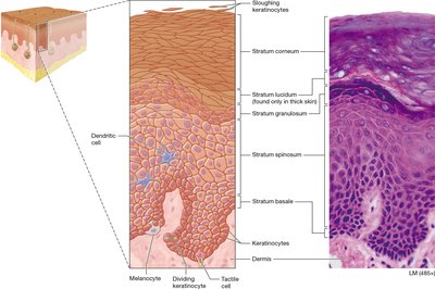
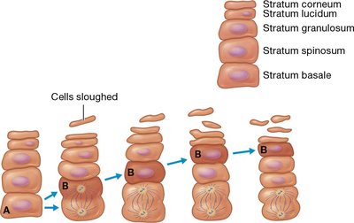
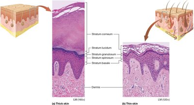
Layers of the Epidermis (Deep to Superficial)
Stratum basale: Single layer of dividing cells; contains keratinocytes, melanocytes, and tactile cells.
Stratum spinosum: Several layers of keratinocytes; contains Langerhans cells.
Stratum granulosum: 3–5 layers; cells flatten, organelles disintegrate, keratinization begins.
Stratum lucidum: Thin, clear layer found only in thick skin (palms, soles).
Stratum corneum: 20–30 layers of dead, keratinized cells; forms a protective barrier.
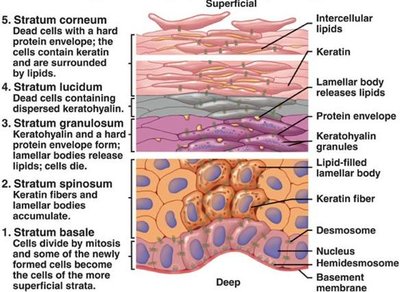
Keratinocyte Life Cycle
Keratinocytes originate in the stratum basale and migrate upward, undergoing differentiation and keratinization.
The entire process from formation to sloughing off at the surface takes 40–50 days.
Thick vs Thin Skin
Thick skin: Contains all five epidermal layers; found on palms, fingertips, and soles; lacks hair and sebaceous glands.
Thin skin: Lacks stratum lucidum; covers most of the body; contains hair and glands.
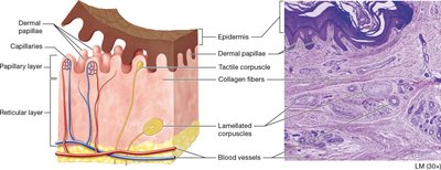
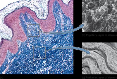
The Dermis
Structure and Layers
Papillary layer: Superficial, areolar connective tissue; contains dermal papillae, capillaries, and tactile corpuscles.
Reticular layer: Deep, dense irregular connective tissue; contains collagen and elastic fibers, blood vessels, glands, and sensory receptors.
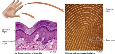
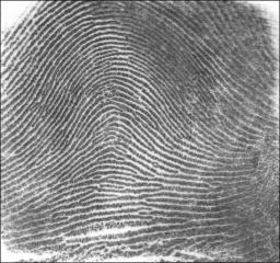
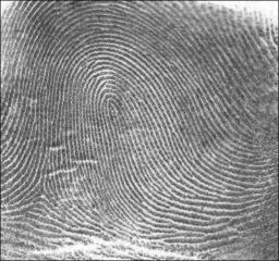
Skin Markings
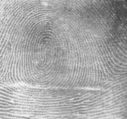
Dermal ridges: Form fingerprints; enhance grip.
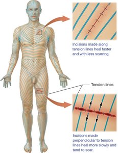
Tension (cleavage) lines: Orientation of collagen bundles; important for surgical incisions.
Flexure lines: Deep creases where the skin is tightly bound to underlying structures, especially around joints.
Skin Pigmentation
Melanin and Other Pigments
Melanin: Produced by melanocytes; protects DNA from UV damage; responsible for skin color variation.
Carotene: Yellow-orange pigment from diet; accumulates in the stratum corneum.
Hemoglobin: Red pigment in blood; gives skin a pinkish hue when dermal blood flow is high.
Clinical Significance of Skin Color
Erythema: Redness due to increased blood flow (e.g., inflammation, sunburn).
Pallor: Paleness from decreased blood flow.
Cyanosis: Bluish color from low oxygen levels.
Jaundice: Yellowing from excess bilirubin (liver dysfunction).
Contusion/Ecchymosis: Bruising from blood vessel disruption.
Accessory Structures: Hair, Nails, and Glands
Hair
Structure: Shaft (above skin), root (below skin), bulb (base), medulla (core), cortex (bulk), cuticle (outer layer).
Types: Lanugo (fetal), vellus (fine, unpigmented), terminal (thick, pigmented).
Functions: Protection, sensation, thermoregulation.
Growth: Cyclic phases—growth, regression, rest.
Nails
Structure: Nail plate (free edge, body, root), nail bed, hyponychium, nail matrix, cuticle (eponychium).
Functions: Protection, enhanced sensation, diagnostic value.
Glands
Sweat glands: Eccrine (thermoregulation), apocrine (axillae, areola, protein-rich), ceruminous (earwax), mammary (milk).
Sebaceous glands: Secrete sebum (oily, hydrophobic, antibacterial); holocrine secretion; associated with hair follicles.
Pathology of the Skin
Wounds and Burns
Wound types: Abrasion, puncture, incision, laceration, avulsion, amputation.
Burn classification:
First-degree: Epidermis only; red, painful.
Second-degree: Epidermis and upper dermis; blisters, severe pain.
Third-degree: Full thickness; destroys entire skin; gray/black, little pain due to nerve damage.
Extent estimation: Rule of Nines (adults), Rule of Eights (infants), % body surface area (BSA).
Burn care: Fluid resuscitation (Parkland Formula): in first 24 hours.
Skin grafts: Autograft (self), allograft (human donor), xenograft (animal donor).
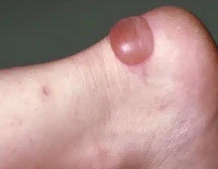
Skin Cancers
Basal cell carcinoma: Most common, least malignant; arises from stratum basale; slow-growing, rarely metastasizes.
Squamous cell carcinoma: From keratinocytes of stratum spinosum; can metastasize if untreated.
Malignant melanoma: From melanocytes; highly metastatic; poor prognosis if not caught early.
ABCDEs of melanoma: Asymmetry, Border irregularity, Color variation, Diameter >6mm, Evolving.
Skin Cancer Type | Origin | Appearance | Prognosis |
|---|---|---|---|
Basal Cell Carcinoma | Stratum basale | Shiny bump, central depression | Excellent if removed early |
Squamous Cell Carcinoma | Stratum spinosum | Red, scaly, ulcerated lesion | Good if removed early |
Malignant Melanoma | Melanocytes | Dark, irregular mole | Poor if metastasized |
