 Back
BackThe Integumentary System: Structure, Function, and Clinical Relevance
Study Guide - Smart Notes
 Tailored notes based on your materials, expanded with key definitions, examples, and context.
The Integumentary System
Overview and Structure
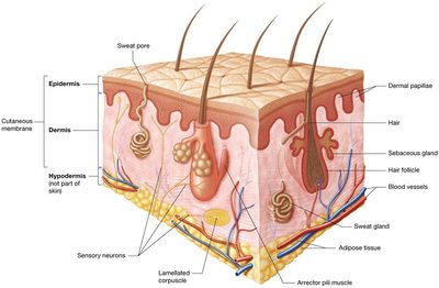
The integumentary system is the largest organ system in the human body, accounting for 10–15% of total body weight. It serves as more than just an outer covering, playing critical roles in protection, sensation, thermoregulation, and homeostasis. The skin, or cutaneous membrane, consists of two main layers: the epidermis and the dermis. Accessory structures such as sweat glands, sebaceous glands, hair, and nails are embedded within the skin, and sensory receptors are distributed throughout.
Epidermis: The superficial layer, composed of keratinized stratified squamous epithelium resting on a basement membrane. It is avascular and relies on diffusion from the dermis for nutrients and oxygen.
Dermis: Located deep to the epidermis, composed of loose connective tissue and dense irregular connective tissue. It is highly vascular and provides structural support and nourishment to the epidermis.
Hypodermis (Superficial Fascia): Not part of the skin proper, but anchors the skin to deeper structures such as muscle and bone. It consists of loose connective and adipose tissues and contains abundant blood supply.
Functions of the Integumentary System
The integumentary system is essential for maintaining homeostasis and protecting the body from external threats.
Protection: The keratinized epithelium protects against mechanical trauma, pathogens, and environmental hazards such as UV radiation. Sebaceous gland secretions create an acidic surface (acid mantle) that inhibits pathogen growth. Lipid-based chemicals provide a hydrophobic barrier, preventing water and electrolyte loss or entry.
Sensation: Numerous sensory receptors detect changes in the internal and external environment, including temperature, pain, and touch.
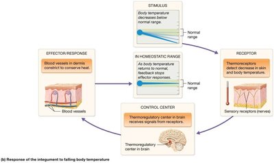
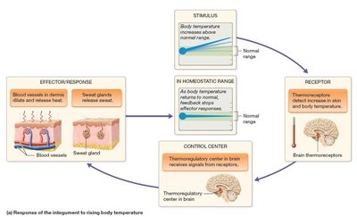
Thermoregulation: The skin helps regulate body temperature through negative feedback loops involving sweating, vasodilation, and vasoconstriction. The hypothalamus acts as the thermoregulatory center.
Excretion: The skin eliminates waste products and toxins through sweat.
Vitamin D Synthesis: The epidermis is involved in the synthesis of vitamin D when exposed to UV light, which is essential for calcium absorption.
The Epidermis
Cell Types and Layers
The epidermis is the most superficial layer of the skin and is composed of several specialized cell types and distinct layers (strata):
Keratinocytes: Make up about 95% of the epidermis. They manufacture keratin, a tough fibrous protein, and are linked by desmosomes.
Dendritic (Langerhans) Cells: Located in the stratum spinosum; act as phagocytes of the immune system.
Merkel Cells: Found in the stratum basale; function as sensory receptors for light touch.
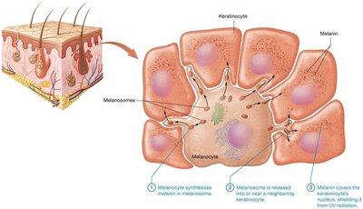
Melanocytes: Also in the stratum basale; produce melanin, a pigment that protects against UV radiation.

Layers of the Epidermis
Stratum Basale (Germinativum): Single layer of stem cells; most metabolically and mitotically active; responsible for vitamin D synthesis and replacement of dead keratinocytes.
Stratum Spinosum: Thickest layer; metabolically and mitotically active.
Stratum Granulosum: 3–5 layers of cells with keratin bundles and lipid-based granules; provides waterproofing.
Stratum Lucidum: Narrow layer of clear, dead keratinocytes; found only in thick skin (e.g., palms, soles).
Stratum Corneum: Outermost layer; several layers of dead, flattened keratinocytes filled with keratin; cells are sloughed off mechanically.
Mnemonic: "Brilliant Studying Gives Loads of Confidence" (Basale, Spinosum, Granulosum, Lucidum, Corneum)
Keratinocyte Life Cycle
Keratinocytes originate in the stratum basale and migrate upwards, undergoing differentiation and eventually dying as they reach the surface. The entire process takes 40–50 days.

The Dermis
Structure and Layers
The dermis is a highly vascular layer deep to the epidermis, providing structural support, nourishment, and sensory functions. It consists of two main layers:
Papillary Layer: The thinner, superficial layer composed of loose connective tissue. Contains dermal papillae, which anchor the epidermis and contain capillaries and tactile (Meissner) corpuscles for light touch sensation.
Reticular Layer: The deeper, thicker layer composed of dense irregular connective tissue. Contains collagen bundles for strength, elastic fibers for flexibility, and proteoglycans for hydration. Also houses blood vessels, sweat glands, sebaceous glands, hair follicles, and sensory receptors (lamellated corpuscles).

Skin Markings and Wrinkles
Dermal Ridges: Areas where dermal papillae are more prominent, forming epidermal ridges (fingerprints) that enhance grip. Patterns are genetically determined and unique to each individual.
Wrinkles: Result from decreased collagen, elastic fibers, proteoglycans, and adipose tissue with age. UV exposure and smoking accelerate wrinkle formation. Treatments include Botox, fillers, and topical creams, though effectiveness varies.

Skin Pigmentation
Melanin and Other Pigments
Skin color is primarily determined by melanin, produced by melanocytes in the stratum basale. Melanin protects keratinocyte DNA from UV-induced mutations and must be continuously produced. Its synthesis increases with UV exposure, leading to tanning. Melanin also reduces vitamin D synthesis, influencing calcium homeostasis.
Melanin: Two types (eumelanin and pheomelanin) produced from tyrosine via tyrosinase. Amount and type of melanin, not the number of melanocytes, determine skin color.
Carotene: Yellow-orange pigment from diet; accumulates in the stratum corneum.
Hemoglobin: Red pigment in blood; imparts pinkish hue to skin depending on blood flow.
Clinical Correlations
Freckles: Localized increased melanin production.
Moles (Nevi): Local proliferation of melanocytes.
Albinism: Genetic lack of tyrosinase, resulting in little or no melanin production and increased risk of UV damage.
Erythema: Redness due to increased blood flow (e.g., fever, infection).
Pallor: Paleness due to decreased blood flow (e.g., cold, anemia).
Cyanosis: Bluish color due to low oxygenation of hemoglobin.
Accessory Structures: Hair, Nails, and Glands
Hair
Hair is composed of keratinized epithelial cells and serves protective and sensory functions. It is found on most body surfaces except thick skin, lips, and parts of the external genitalia.
Shaft: Visible part above the skin surface; composed of dead, keratinized cells.
Root: Embedded in the dermis; contains living cells and is surrounded by the hair follicle.
Hair Bulb: Base of the root, containing the hair papilla (blood supply) and matrix (growth region).
Arrector Pili Muscle: Smooth muscle that causes hair to stand up (goosebumps) when contracted.

Hair Types and Pigmentation
Lanugo: Thin, nonpigmented hair on fetuses.
Terminal Hair: Thick, pigmented hair on scalp, eyebrows, and after puberty in certain regions.
Vellus Hair: Thin, nonpigmented hair covering most of the body.
Hair Color: Determined by type and amount of melanin; decreases with age, leading to gray or white hair.
Glands
Sudoriferous (Sweat) Glands: Produce sweat for thermoregulation and excretion. Types include eccrine (widely distributed, watery secretion) and apocrine (axillae, anal region, areolae; thicker secretion influenced by hormones).
Sebaceous Glands: Secrete oily sebum onto hair follicles; provides hydrophobic barrier and inhibits bacterial growth.
Ceruminous Glands: Modified apocrine glands in the ear; produce cerumen (ear wax) for protection and lubrication.
Mammary Glands: Specialized sweat glands that produce milk.
Additional info: The integumentary system is also involved in immune defense, sensory perception, and plays a role in social and sexual communication through skin appearance and scent.
