 Back
BackThe Integumentary System: Structure, Function, and Layers
Study Guide - Smart Notes
 Tailored notes based on your materials, expanded with key definitions, examples, and context.
The Integumentary System
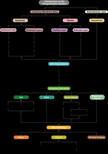
Introduction to the Integumentary System
The integumentary system is an organ system that includes the skin, hair, nails, glands, and sensory receptors. It is the body's largest organ system and serves as the primary interface between the body and the external environment. The system is composed of three main parts: the epidermis, dermis, and the hypodermis (subcutaneous layer), each with distinct structures and functions.
Cutaneous membrane (skin): Consists of the epidermis and dermis.
Accessory structures: Includes hair, nails, and glands (sweat and sebaceous).
Hypodermis: Lies beneath the cutaneous membrane and anchors the skin to underlying tissues.
Functions of the Integumentary System
The integumentary system performs several vital functions essential for maintaining homeostasis and protecting the body:
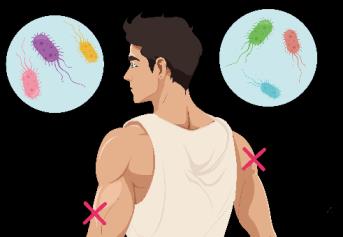
Protection: Acts as a barrier against mechanical injury, chemicals, UV radiation, and pathogens.
Thermoregulation: Regulates body temperature through vasodilation, vasoconstriction, and sweating.
Sensation: Contains sensory receptors for touch, pain, temperature, and pressure.
Vitamin D synthesis: Initiates the production of vitamin D when exposed to sunlight.
Excretion: Removes waste products via sweat.
Communication: Allows for expressive communication through facial expressions and skin color changes.
Organization of the Integumentary System
The system is organized into layers and accessory structures, each contributing to its overall function.
Thermoregulation
Mechanisms of Thermoregulation
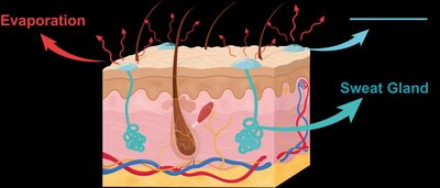
The integumentary system maintains a stable internal body temperature through two main mechanisms: vasoconstriction/vasodilation and sweating.
Vasoconstriction: Blood vessels constrict (decrease in diameter) when the body is cold, reducing blood flow to the skin and minimizing heat loss.
Vasodilation: Blood vessels dilate (increase in diameter) when the body is hot, increasing blood flow to the skin and facilitating heat loss.
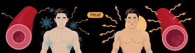
Sweating: Sweat glands secrete a water-based solution onto the skin surface. As sweat evaporates, it cools the body.
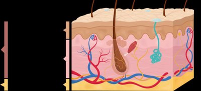
The Epidermis
Cell Types in the Epidermis
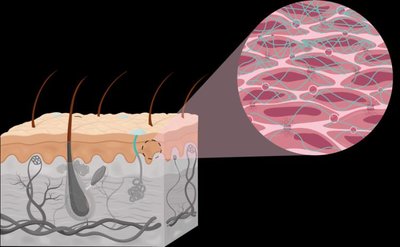
The epidermis is the outermost layer of the skin, composed of stratified squamous epithelial tissue. It contains four main cell types:
Keratinocytes: Most abundant; produce keratin, a tough, water-resistant protein that provides mechanical strength and protection.
Melanocytes: Produce melanin, a pigment that protects against UV damage.
Dendritic (Langerhans) cells: Immune cells that help initiate immune responses.
Tactile epithelial (Merkel) cells: Specialized for touch sensation.
Layers of the Epidermis
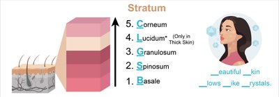
The epidermis is organized into distinct layers (strata), each with specific characteristics and functions. From deep to superficial, the layers are:
Stratum basale: Single row of proliferating cells; contains keratinocytes, melanocytes, and tactile cells.
Stratum spinosum: Several layers of keratinocytes; contains dendritic cells for immunity.
Stratum granulosum: Keratinocytes stop dividing, begin to harden, and accumulate keratin; cells start to die.
Stratum lucidum: Present only in thick skin (palms, soles); clear, dead, densely packed cells.
Stratum corneum: Outermost layer; dead, keratin-filled cells that are regularly shed and replaced.
Thin vs. Thick Skin
Feature | Thin Skin | Thick Skin |
|---|---|---|
Stratum lucidum | Absent | Present |
Location | Most of body | Palms, soles |
Hair follicles & oil glands | Present | Absent |
Sweat glands | Fewer | More numerous |
Keratinocyte Development
Keratinocytes originate in the stratum basale and are gradually pushed toward the surface, undergoing changes as they move through each layer. By the time they reach the stratum corneum, they are dead, flattened, and filled with keratin.

The Dermis
Structure and Layers of the Dermis
The dermis is the second layer of the skin, located beneath the epidermis. It is composed of two layers:
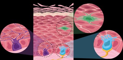
Papillary layer: Superficial; made of loose areolar connective tissue. Contains capillaries, lymphatics, and Meissner corpuscles (touch receptors). Dermal papillae create surface ridges (fingerprints).
Reticular layer: Deep; made of dense irregular connective tissue. Contains sweat and oil glands, hair roots, and Pacinian corpuscles (pressure receptors). Collagen and elastic fibers provide strength and flexibility; cleavage lines are important for surgical incisions.
The Hypodermis (Subcutaneous Layer)
Structure and Function
The hypodermis is a layer of adipose and areolar connective tissue located beneath the dermis. It is not technically part of the skin but serves important roles:
Anchors the skin to underlying tissues.
Acts as a shock absorber to protect internal organs.
Serves as an insulator to reduce heat loss.
Stores energy in the form of fat.

Summary Table: Layers of the Skin
Layer | Main Tissue | Key Features |
|---|---|---|
Epidermis | Stratified squamous epithelium | Protection, keratinocytes, avascular |
Dermis (Papillary) | Areolar connective tissue | Capillaries, touch receptors, dermal papillae |
Dermis (Reticular) | Dense irregular connective tissue | Strength, flexibility, glands, hair roots |
Hypodermis | Adipose & areolar tissue | Anchoring, insulation, fat storage |
Additional info: The integumentary system is essential for homeostasis, immune defense, and sensory perception. Disorders of the skin can impact overall health and are often indicators of systemic disease.
