Back
BackThe Integumentary System: Structure, Function, and Clinical Aspects
Study Guide - Smart Notes
 Tailored notes based on your materials, expanded with key definitions, examples, and context.
The Integumentary System
Overview of the Skin
The integumentary system is composed of the skin and its derivatives, including hair, nails, and glands. The skin is the largest organ of the body, serving as a protective barrier and performing multiple physiological functions.
Surface Area: 1.5–2 square meters
Weight: 4–5 kg
Thickness: Varies from 1.5 mm to 4 mm
Functions of the Skin
Regulation of Body Temperature: Acts as an insulator and radiator to maintain homeostasis.
Protection: Physical barrier against mechanical injury, pathogens, and water loss.
Sensation: Contains sensory nerve endings for touch, pain, temperature, and pressure.
Communication: Facilitates nonverbal communication through facial expressions and touch.
Excretion: Minor role in excreting water, salts, and small organic compounds via sweat.
Immunity: Epidermal phagocytes contribute to immune defense.
Blood Reservoir: The dermis contains a significant portion of the body's blood supply.
Synthesis of Vitamin D: UV light triggers vitamin D production, essential for calcium absorption.
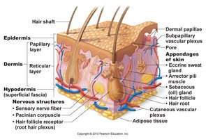
Skin Structure
Principal Layers of the Skin
The skin consists of three main layers: the epidermis, dermis, and hypodermis (subcutaneous layer). Each layer has distinct structural and functional properties.
Epidermis: Stratified squamous epithelium; provides a waterproof barrier and creates skin tone.
Dermis: Areolar and dense irregular connective tissue; provides strength, elasticity, and houses blood vessels, nerves, and glands.
Hypodermis: Subcutaneous layer composed mainly of adipose tissue; anchors skin to underlying tissues.
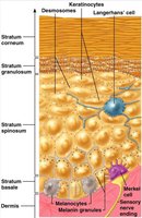
The Epidermis: Cell Types and Layers
The epidermis is composed of four main cell types and organized into several layers, each with specialized functions.
Keratinocytes (90%): Produce keratin, a protein that provides strength and waterproofing.
Melanocytes (8%): Synthesize melanin pigment, which protects against UV radiation.
Langerhans Cells: Immune cells involved in defense against pathogens.
Merkel Cells: Sensory receptors for touch, located in the deepest layer of hairless skin.

Epidermal Layers
Stratum Basale: Single layer of cuboidal/columnar cells; contains stem cells, melanocytes, and Merkel cells. Site of cell division (mitosis).
Stratum Spinosum: 8–10 layers of keratinocytes; contains Langerhans cells and desmosomes for cell adhesion.
Stratum Granulosum: 3–5 layers of flattened cells with keratohyaline granules; initiation of cell death.
Stratum Lucidum: Present only in thick skin (palms, soles); 3–5 layers of clear, dead cells.
Stratum Corneum: 20–30 layers of dead, keratin-filled cells; continuously shed and replaced.
The Dermis
The dermis provides structural support and elasticity to the skin. It is divided into two regions:
Papillary Region (20%): Areolar connective tissue with dermal papillae that increase surface area for nutrient exchange; contains Meissner's corpuscles for light touch.
Reticular Region (80%): Dense irregular connective tissue with collagen and elastic fibers; provides strength and extensibility. Tears in this region cause stretch marks.

Skin Pigmentation
Major Skin Pigments
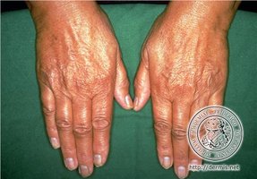
Melanin: Yellow to black pigment produced by melanocytes; protects against UV radiation. The amount and type of melanin varies among individuals.
Carotene: Yellow-orange pigment stored in the dermis and subcutaneous layer; precursor for vitamin A.
Hemoglobin: Red pigment in blood; gives skin a pinkish hue, especially in lighter-skinned individuals.
Clinical Terminology of Skin Color
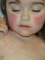
Erythema: Redness due to increased blood flow (e.g., exercise, inflammation).
Pallor: Paleness from reduced blood flow or anemia.
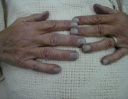
Cyanosis: Bluish tint from lack of oxygen in the blood.
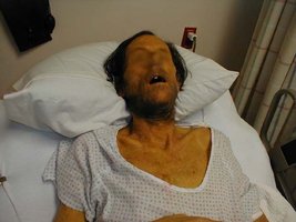
Jaundice: Yellow/orange color from bilirubin accumulation, often due to liver dysfunction.
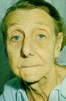
Bronzing: Metallic appearance, seen in Addison's disease.
Contusions: Black and blue marks (bruises) from blood leakage under the skin.
Skin Pathologies
Skin Cancers
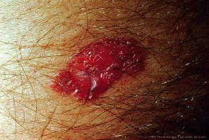
Basal Cell Carcinoma: Originates from the stratum basale; least malignant, high cure rate.
Squamous Cell Carcinoma: Arises from the stratum spinosum; good prognosis if treated early.
Melanoma: Cancer of melanocytes; highly metastatic and resistant to chemotherapy.
ABCD Rule for Melanoma Detection:
Asymmetry
Border irregularity
Color variation
Diameter > 6 mm
Melanin Pathologies
Albinism: Genetic disorder preventing melanin synthesis; results in very light skin, hair, and eyes.
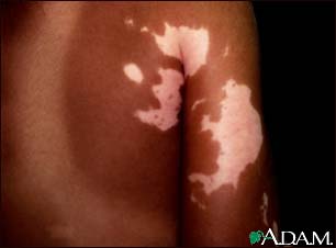
Vitiligo: Loss of melanocytes in patches, causing depigmented areas.

Burns
Classification of Burns
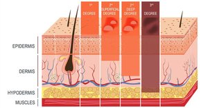
First Degree: Only epidermis is damaged; redness, swelling, pain; heals in 2–3 days.
Second Degree: Epidermis and upper dermis damaged; blisters form; heals in 3–4 weeks.
Third Degree: Entire thickness of skin destroyed; area appears gray-white, cherry red, or black; no initial pain due to nerve destruction; requires skin grafting.
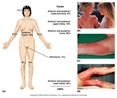
Rule of Nines
The Rule of Nines is used to estimate the percentage of body surface area affected by burns, guiding treatment decisions.
Burns are critical if >25% of body has second-degree burns, >10% has third-degree burns, or if third-degree burns affect face, hands, or feet.
Skin Grafts
Autograft: Skin from the same person
Isograft: Skin from an identical twin
Homograft: Skin from another human
Heterograft: Skin from an animal
Epidermal Derivatives
Hair (Pili)
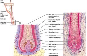
Hair serves protective, sensory, and social functions. Its structure includes the shaft, root, follicle, and bulb.
Shaft: Visible part; composed of medulla, cortex, and cuticle.
Root: Portion within the skin; similar structure to shaft.
Follicle: Surrounds the root; includes a sheath and bulb at the base.
Bulb: Contains the papilla (blood supply) and matrix (growth zone).
Sebaceous Glands: Release oil into the follicle.
Arrector Pili: Smooth muscle causing hair to stand (goosebumps).
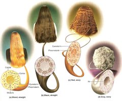
Hair Color and Growth
Brown/Black: Melanin deposited in shaft.
Red/Blond: Variants of melanin with iron and sulfur.
Gray: Reduced melanin due to loss of melanocytes.
White: Air bubbles in cortex.
Hair and Hormones
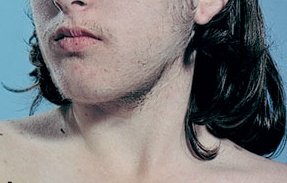
Testosterone: Increases at puberty, influencing hair growth patterns.
Hirsutism: Excess hair growth in females or prepubertal males due to elevated testosterone.
Male Pattern Baldness: Genetic, sex-linked trait; influenced by dihydrotestosterone (DHT).


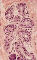
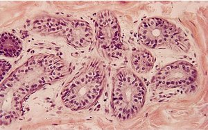
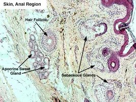
Glands of the Skin
Sebaceous (Oil) Glands
Connected to hair follicles; secrete sebum (oil) to lubricate hair and skin.
Holocrine glands; secretion includes fats, cholesterol, proteins, and salts.
Prevents water loss and inhibits bacterial growth.
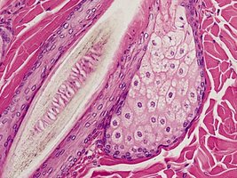
Sudoriferous (Sweat) Glands
Eccrine Glands: Most abundant; regulate body temperature; found on palms, soles, and forehead.
Apocrine Glands: Located in axillary, anogenital regions, and areolae; secrete during stress and sexual arousal.
Modified Sweat Glands
Ceruminous Glands: Produce earwax (cerumen) for protection.
Mammary Glands: Specialized for milk production; regulated by hormones.
Cystic Fibrosis and Sweat Glands
Autosomal recessive disorder affecting the CFTR gene.
Results in abnormal chloride transport and thick glandular secretions.
Diagnosed by elevated chloride in sweat.
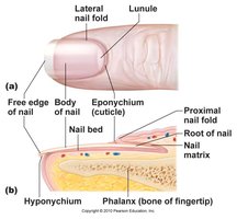
Nails
Structure and Growth
Composed of tightly packed, hard keratinized cells.
Nail Matrix: Site of nail growth; transforms skin cells into nail cells.
Growth rate: ~1 mm/week in fingers; slower in toes.