 Back
BackThe Integumentary System: Structure, Function, and Clinical Aspects
Study Guide - Smart Notes
 Tailored notes based on your materials, expanded with key definitions, examples, and context.
Chapter 5: The Integumentary System
Overview of the Integumentary System
The integumentary system is the largest organ system in the human body, comprising the skin and its accessory structures. It serves as the primary barrier between the internal environment and the external world, playing crucial roles in protection, sensation, thermoregulation, and metabolic functions.
Major Components: Skin tissue and accessory structures (exocrine glands, hair, nails)
Surface Area: Approximately 1.5–2 m2
Body Weight: Accounts for about 16% of total body weight
Functions of the Integumentary System:
Protection of underlying tissues and organs
Excretion of wastes and regulation of body temperature via sweating
Production of melanin (UV protection) and keratin (water/abrasion resistance)
Synthesis of vitamin D3 for bone development and homeostasis
Detection of sensations (touch, pressure, pain, temperature, vibration)
Coordination of immune responses
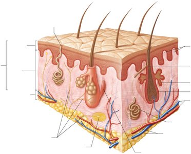
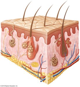
Skin Structure
Layers of the Skin
The skin consists of two main layers: the epidermis and the dermis. Beneath these lies the hypodermis (subcutaneous layer), which is not technically part of the skin but supports its functions.
Epidermis: Outermost, avascular layer composed of stratified squamous epithelium
Dermis: Deeper, vascular layer containing connective tissue, blood vessels, nerves, and accessory structures
Hypodermis: Subcutaneous tissue primarily made of adipose tissue; stabilizes skin and stores energy
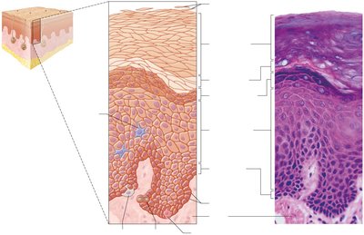
Epidermal Layers (Strata)
The epidermis is organized into distinct layers, each with specialized cells and functions. The number of layers differs between thin and thick skin.
Stratum Corneum: Multiple layers of dead, keratinized cells; water-resistant
Stratum Lucidum: Thin, clear layer found only in thick skin (palms, soles)
Stratum Granulosum: Keratinocytes produce keratin and begin to die
Stratum Spinosum: Keratinocytes bound by desmosomes; contains dendritic cells
Stratum Basale: Single layer of stem cells; site of cell division and melanocytes
Thick Skin vs. Thin Skin:
Thick Skin: Five layers; found on palms and soles
Thin Skin: Four layers; covers most of the body
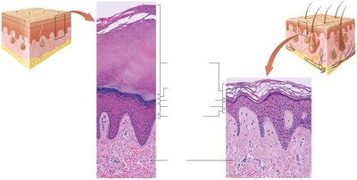
Dermis Structure
The dermis is divided into two layers, each with distinct connective tissue types and functions.
Papillary Layer: Superficial; areolar connective tissue, contains capillaries, lymphatics, and sensory neurons
Reticular Layer: Deep; dense irregular connective tissue with collagen and elastic fibers, provides strength and elasticity

Hypodermis (Subcutaneous Layer)
The hypodermis lies beneath the dermis and is primarily composed of adipose tissue. It is not considered part of the integumentary system but plays a role in insulation, energy storage, and anchoring the skin to underlying tissues.
Contains large blood vessels and nerves
Distribution of fat varies by sex hormones
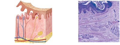
Accessory Structures of the Skin
Exocrine Glands
Accessory structures such as glands, hair, and nails are derived from the epidermis and play specialized roles in skin function.
Merocrine (Eccrine) Glands: Secrete watery sweat for thermoregulation and waste excretion
Apocrine Glands: Release a portion of cytoplasm, causing body odor; active after puberty
Holocrine Glands (Sebaceous): Discharge sebum (oil) into hair follicles; lubricates and protects skin


Skin Pigmentation and Clinical Correlates
Melanin and Skin Color
Melanin, produced by melanocytes in the stratum basale, is the primary pigment responsible for skin color. Melanin protects keratinocyte DNA from UV radiation by forming a protective cap over the nucleus.
Melanosomes: Organelles that synthesize and transport melanin to keratinocytes
Other Pigments: Carotene (orange pigment), hemoglobin (redness from blood flow)
Skin Color and Disease
Changes in skin color can indicate underlying health conditions:
Cyanosis: Blue tint due to low oxygen
Erythema: Redness from increased blood flow
Jaundice: Yellowing from bile accumulation (liver dysfunction)
Pituitary Tumor/Addison’s Disease: Excess melanin production
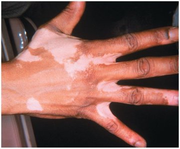
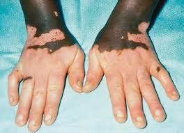
Vitiligo: Loss of melanocytes, resulting in patchy depigmentation
Vitamin D3 Synthesis
Vitamin D3 is synthesized in the epidermis upon exposure to UV radiation. It is converted by the liver and kidneys into calcitriol, which is essential for calcium absorption and bone health. Deficiency can lead to rickets.
Skin Damage and Repair
Types of Skin Damage
Loss of Skin Turgor: Caused by dehydration, aging, or hormonal changes
Stretch Marks: Result from excessive stretching (e.g., pregnancy, weight gain)
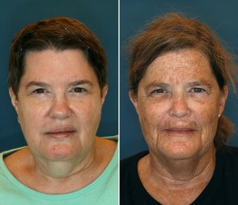
Photoaging: Sunlight accelerates aging by damaging collagen and elastic fibers
UV Radiation: Can cause DNA damage and increase risk of skin cancer
Skin Cancer
There are three major types of skin cancer, each with distinct characteristics and risks:
Type | Origin | Appearance | Risk | Treatment |
|---|---|---|---|---|
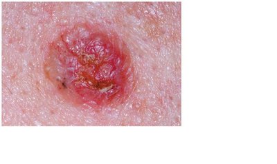
Basal Cell Carcinoma | Stratum basale | Shiny elevation, central depression, pearly edge | Least dangerous, rarely metastasizes | Surgical removal |
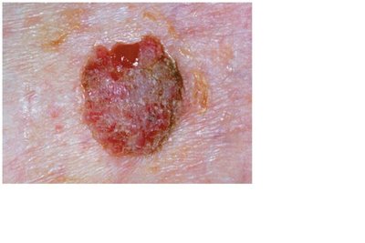
Squamous Cell Carcinoma | Stratum spinosum | Raised, scaly, later ulcerated | May metastasize | Early detection, surgical removal |
Malignant Melanoma | Melanocytes (often in moles) | Asymmetry, irregular border, color variation, diameter >6mm, evolving | Most deadly, aggressive metastasis | Early removal, chemotherapy, radiation |
ABCDE Rule for Melanoma Detection:
Asymmetry
Border irregularity
Color variation
Diameter > 6 mm
Evolving shape, size, or symptoms
Burns and Classification
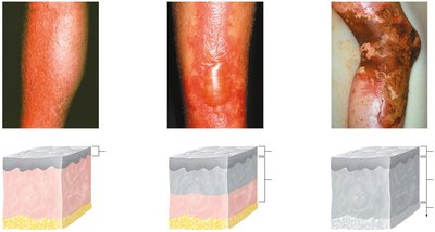
Burns are classified by depth and extent of tissue damage:
First-degree: Involves only the epidermis (redness, pain)
Second-degree: Involves epidermis and part/all of dermis (blisters, swelling)
Third-degree: Involves epidermis, dermis, hypodermis, and possibly deeper tissues (charred, insensate)
The Rule of Nines is used to estimate the extent of burns for clinical management.
Tissue Repair
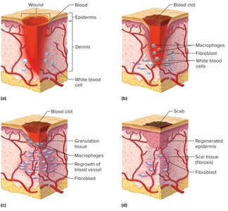
Skin repair involves several stages: inflammation, migration of cells, proliferation, and maturation. Fibroblasts produce scar tissue, and the epidermis regenerates over time.
Additional info: The integumentary system is essential for homeostasis, immune defense, and sensory perception. Disorders of the skin can reflect systemic diseases and require interdisciplinary management.
