Back
BackThe Integumentary System: Structure, Function, and Clinical Aspects
Study Guide - Smart Notes
 Tailored notes based on your materials, expanded with key definitions, examples, and context.
The Integumentary System
Overview
The integumentary system is the body's outer covering, providing protection and contributing to homeostasis. It consists of the skin, hair, nails, sweat glands, and sebaceous (oil) glands. The skin is the largest organ and serves as a barrier against environmental hazards.
Major components: Skin, hair, nails, sweat glands, sebaceous glands
Main functions: Protection, sensation, temperature regulation, metabolic functions, excretion
Structure of the Skin
Skin Layers
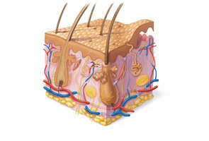
The skin is composed of two main layers: the epidermis and the dermis. Beneath these lies the hypodermis (subcutaneous tissue), which is not technically part of the skin but supports its functions.
Epidermis: Superficial, avascular layer made of keratinized stratified squamous epithelium
Dermis: Deeper, vascular layer composed of connective tissue
Hypodermis: Subcutaneous layer of adipose tissue; absorbs shock, insulates, and anchors skin to muscles
Cells of the Epidermis
Major Cell Types
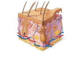
The epidermis contains four main cell types, each with specialized functions:
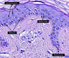
Keratinocytes: Produce keratin, a protein that provides strength and waterproofing; most abundant cell type
Melanocytes: Produce melanin pigment, which protects against UV radiation
Dendritic (Langerhans) cells: Immune cells that patrol the epidermis and activate immune responses
Tactile (Merkel) cells: Sensory receptors for touch
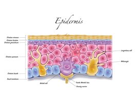
Layers of the Epidermis
Strata of the Epidermis
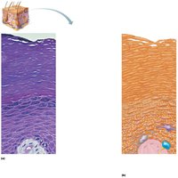
The epidermis is organized into distinct layers (strata). Thick skin (palms, soles) has five layers; thin skin has four.
Stratum basale (germinativum): Deepest, single row of mitotic stem cells; contains melanocytes
Stratum spinosum: Several layers of keratinocytes connected by desmosomes; contains dendritic cells and melanosomes
Stratum granulosum: 4–6 layers; keratinization begins, cells flatten, organelles disintegrate, lamellar granules release glycolipids
Stratum lucidum: Only in thick skin; thin, clear layer of dead keratinocytes
Stratum corneum: 20–30 layers of dead, keratinized cells; provides barrier function
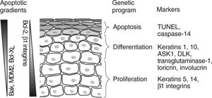
Cell Turnover and Shedding
Epidermal cells undergo apoptosis as they move toward the surface, eventually sloughing off as dandruff and dander. Humans shed approximately 50,000 skin cells per minute.
The Dermis
Structure and Layers
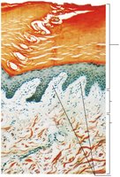
The dermis is a strong, flexible connective tissue layer containing fibroblasts, macrophages, mast cells, and white blood cells. It houses nerves, blood vessels, lymphatics, hair follicles, and glands.
Papillary layer: Superficial areolar connective tissue with dermal papillae (capillary loops, nerve endings, touch receptors)
Reticular layer: Deep, dense irregular connective tissue; provides strength, elasticity, and hydration
Dermal Modifications and Skin Markings
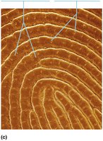
Friction ridges: Form fingerprints; enhance grip and tactile sensitivity
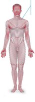
Cleavage (tension) lines: Orientation of collagen fibers; important for surgical incisions
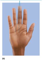
Flexure lines: Dermal folds at joints; allow skin to accommodate movement
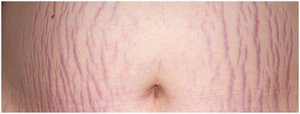
Striae (stretch marks): Dermal tears from overstretching
Skin Color
Pigments
Skin color is determined by three main pigments:
Melanin: Produced by melanocytes; protects DNA from UV damage; responsible for brown to black hues
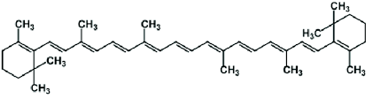
Carotene: Yellow to orange pigment; accumulates in stratum corneum and hypodermis; can be converted to vitamin A
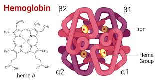
Hemoglobin: Oxygen-carrying pigment in red blood cells; gives pinkish hue to fair skin
Clinical Correlations: Skin Color Changes
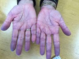
Cyanosis: Blue skin due to low oxygenation
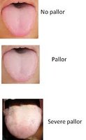
Pallor: Pale color from anemia, low blood pressure, or shock
Erythema: Redness from fever, inflammation, or allergy
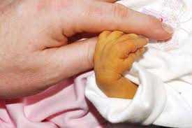
Jaundice: Yellow cast from liver disorders
Bruises: Clotted blood beneath skin (hematomas)
Hyperpigmentation: May indicate endocrine disorders
Hair
Structure and Function
Hair consists of dead, keratinized cells produced by hair follicles. It functions in protection, sensation, and temperature regulation.
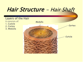
Shaft: Visible part above the skin; keratinization complete
Root: Embedded in the skin; keratinization ongoing
Medulla: Central core with air spaces
Cortex: Layers of flattened cells surrounding the medulla
Cuticle: Outermost layer of overlapping cells
Hair Color
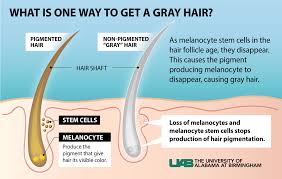
Hair color is determined by the type and amount of melanin produced by melanocytes in the hair follicle. Red hair contains pheomelanin. Gray or white hair results from decreased melanin production and air bubbles in the shaft.
Hair Follicle Structure
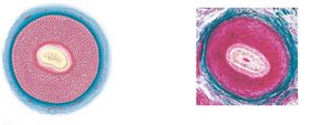
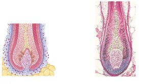
The hair follicle extends from the epidermis into the dermis and consists of several layers:
Peripheral connective tissue sheath: Derived from dermis
Glassy membrane: Thickened basal lamina
Epithelial root sheath: Derived from epidermis
Hair matrix: Actively dividing cells producing hair
Arrector pili: Smooth muscle causing "goose bumps"
Hair papilla: Dermal tissue supplying nutrients
Types and Growth of Hair
Vellus hair: Fine, pale body hair
Terminal hair: Coarse, long hair (scalp, eyebrows, pubic, axillary regions)
Growth: Averages 2.25 mm/week; affected by nutrition and hormones
Hair Thinning and Baldness
Alopecia: Age-related hair thinning
Male pattern baldness: Genetically determined, influenced by DHT
Telogen effluvium: Sudden hair loss due to stress or illness
Nails
Structure and Function
Nails are scale-like modifications of the epidermis containing hard keratin. They protect the distal tips of fingers and toes and aid in manipulation.
Free edge: Extends beyond the finger or toe
Nail plate: Visible attached portion
Nail root: Embedded in the skin
Nail matrix: Site of nail growth
Lunule: White crescent at the base of the nail
Eponychium (cuticle): Skin fold over the nail
Hyponychium: Area under the free edge
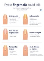
Clinical Correlations: Nail Appearance
Yellow nails: May indicate respiratory or thyroid disorders
Thickened yellow nails: Fungal infection
Spoon nails (koilonychia): Iron deficiency
Beau’s lines: Severe illness
Cutaneous Glands
Types of Glands
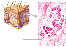
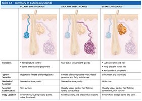
Eccrine (merocrine) sweat glands: Most numerous; thermoregulation; secrete watery sweat
Apocrine sweat glands: Axillary/anogenital areas; secrete viscous sweat; function at puberty
Modified apocrine glands: Ceruminous (earwax) and mammary (milk) glands
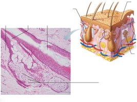
Sebaceous (oil) glands: Secrete sebum into hair follicles; lubricate and protect skin/hair
Gland Type | Function | Secretion | Location |
|---|---|---|---|
Eccrine Sweat Gland | Temperature control, antibacterial | Hypotonic filtrate of blood plasma (mostly water, some salts) | Everywhere, especially palms, soles, forehead |
Apocrine Sweat Gland | May act as sexual scent gland | Filtrate of blood plasma with added proteins and fatty substances | Axillary and anogenital regions |
Sebaceous Gland | Lubricate skin/hair, antibacterial | Oily sebum | Everywhere except palms and soles |
Clinical Correlations: Gland Disorders
Acne: Inflammation of sebaceous glands, often due to bacterial infection
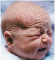
Seborrhea (cradle cap): Overactive sebaceous glands in infants
Functions of the Skin
Protection
Chemical barrier: Sweat, sebum, acid mantle, melanin
Physical barrier: Keratinized cells, glycolipids
Biological barrier: Dendritic cells, macrophages, DNA (UV absorption)
Body Temperature Regulation
Insensible perspiration: Unnoticeable sweat at rest (~500 ml/day)
Sensible perspiration: Noticeable sweat during heat or exercise (up to 12 L/day)
Vasoconstriction: Reduces heat loss in cold environments
Cutaneous Sensations
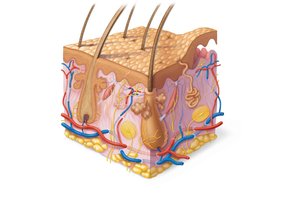
Exteroreceptors: Detect temperature, touch, pain
Free nerve endings: Sense pain
Metabolic Functions
Synthesizes vitamin D for calcium absorption
Detoxifies some chemicals and activates hormones
Makes collagenase for collagen turnover
Blood Reservoir and Excretion
Holds up to 5% of blood volume
Excretes nitrogenous wastes, salt, and water
Skin Cancer and Burns
Skin Cancer
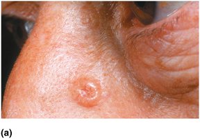
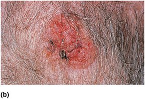
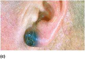
Basal cell carcinoma: Most common, least malignant; arises from stratum basale
Squamous cell carcinoma: Second most common; can metastasize; arises from stratum spinosum
Melanoma: Most dangerous; cancer of melanocytes; highly metastatic
Burns
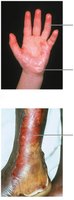
First-degree: Epidermal damage only; redness, pain
Second-degree: Epidermal and upper dermal damage; blisters
Third-degree: Full-thickness; gray-white, cherry red, or blackened; no pain (nerve endings destroyed); requires grafting
Rule of Nines
The Rule of Nines is used to estimate the extent of burns and guide fluid resuscitation. The body is divided into sections, each accounting for 9% (or multiples) of total body surface area.
Burns are critical if: >25% of body has second-degree burns, >10% has third-degree burns, or face/hands/feet have third-degree burns
Treatment includes debridement, antibiotics, temporary coverings, and skin grafts