Back
BackThe Integumentary System: Structure, Function, and Clinical Relevance
Study Guide - Smart Notes
 Tailored notes based on your materials, expanded with key definitions, examples, and context.
The Integumentary System
Introduction to the Integumentary System
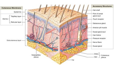
The integumentary system is the body's first line of defense, providing a protective barrier against environmental hazards. It consists of the cutaneous membrane (skin) and accessory structures such as hair, nails, muscles, sensory receptors, and glands. The skin is the largest organ in the body by weight.
Protection: Prevents water loss and entry of pathogens.
Thermoregulation: Maintains body temperature through sweat and blood flow.
Synthesis and Storage of Nutrients: Produces vitamin D3 and stores lipids.
Sensory Reception: Detects touch, pressure, pain, and temperature.
Excretion and Secretion: Removes waste products via sweat and sebum.
Skin Structure
Layers of the Skin
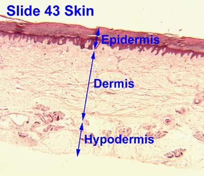
The skin is composed of two main layers: the epidermis and the dermis. Beneath these lies the hypodermis (subcutaneous layer), which is not technically part of the skin but is important for fat storage and insulation.
Epidermis: Stratified squamous epithelium; avascular; contains keratinocytes.
Dermis: Areolar connective tissue (papillary layer) and dense irregular connective tissue (reticular layer); provides blood supply and structural strength.
Hypodermis: Composed mainly of adipose tissue with some areolar connective tissue; site for subcutaneous injections.
Definition of an Organ
An organ is a structure composed of at least two tissue types that performs a specific function for the body.
Microscopic Structure of the Skin
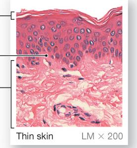
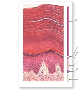
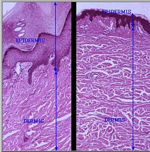
Histological slides reveal the organization of the epidermis and dermis, as well as the differences between thin and thick skin.
Epidermis
Characteristics and Functions
The epidermis is avascular and consists of stratified squamous epithelium. Its main cell type is the keratinocyte, which produces keratin for protection against water loss, injury, chemicals, and microorganisms.
Five Layers (Strata) in Thick Skin (from deep to superficial):
Stratum basale: Single layer of stem cells; contains melanocytes and Merkel cells.
Stratum spinosum: Several layers; contains dendritic cells for immune defense.
Stratum granulosum: Cells stop dividing and begin keratinization.
Stratum lucidum: Present only in thick skin; cells are dead and clear.
Stratum corneum: Most superficial; layers of dead, keratinized cells.
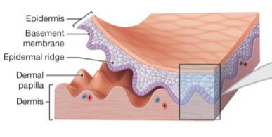
Epidermal Ridges and Dermal Papillae: Increase surface area for attachment and nutrient exchange.
Thick vs. Thin Skin
Thin Skin: Covers most of the body; four strata; ~0.08 mm thick.
Thick Skin: Found on palms and soles; five strata; ~0.5 mm thick.
Skin Pigmentation
Types of Pigments
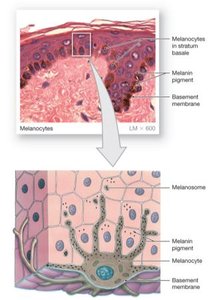
Skin color is determined by the presence of carotene (from diet) and melanin (produced by melanocytes). Melanocytes are located in the deepest epidermal layer and the dermis. UV exposure increases melanin production, which protects against DNA damage.
Genetics and Environment: All humans have similar numbers of melanocytes, but pigment production and metabolism vary.
Vitamin D3 Synthesis
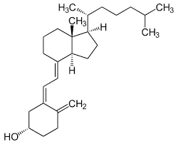
Vitamin D3 is synthesized by epidermal cells when exposed to UV radiation. It is converted by the liver and kidneys into calcitriol, which is essential for calcium and phosphorus absorption. Deficiency can lead to weak bones (rickets).
Skin Cancer
Types and Risk Factors
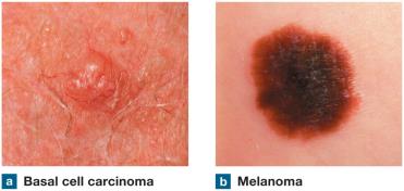
Excessive sun exposure increases the risk of skin cancer. The most common types are basal cell carcinoma and squamous cell carcinoma, which are usually benign. Malignant melanoma is more dangerous due to its ability to metastasize.
ABC's of Skin Cancer:
A: Asymmetry
B: Border irregularity
C: Color variation
D: Diameter > 6mm
E: Elevation
The Dermis
Structure and Function
The dermis lies beneath the epidermis and provides structural support, elasticity, and houses blood vessels, glands, hair follicles, and nerves. It consists of two layers:
Papillary Layer: Areolar connective tissue; forms dermal papillae.
Reticular Layer: Dense irregular connective tissue; provides strength and elasticity.
Collagen Fibers, Tension Lines, and Wrinkles
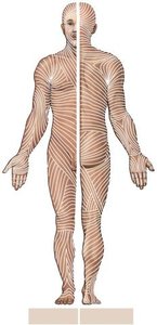
Collagen fibers in the dermis are oriented in tension lines, which are important for surgical incisions and healing. Wrinkles and sagging occur when collagen is damaged or replaced slowly with age.
Functions of the Integument
Protection, Sensation, and Homeostasis
Melanin: Protects against UV radiation.
Keratin: Provides waterproofing and mechanical protection.
Immune Responses: Dendritic cells in the epidermis detect pathogens.
Sensory Nerve Endings: Detect environmental changes.
Lipid Storage: Hypodermis stores energy as fat.
Vitamin D Production: Essential for bone health.
Temperature Regulation: Achieved through sweat and blood flow.

Temperature Regulation
Heat is produced by metabolic reactions and distributed by blood. The skin responds to increased temperature by sweating and vasodilation, and to decreased temperature by vasoconstriction and shivering.
Accessory Structures
Hair
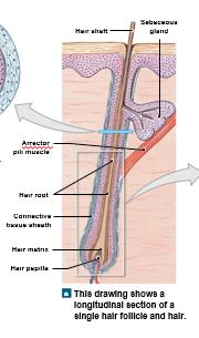
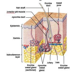
Hair is composed of dead, keratinized epithelial cells. It provides protection, insulation, and sensory input. The arrector pili muscle causes hair to stand up (goosebumps).
Nails
Nails are made of dead, keratinized epidermal cells and protect the tips of fingers and toes.
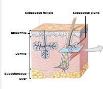
Sebaceous (Oil) Glands
These glands are associated with hair follicles and secrete sebum, which softens hair and skin and has antibacterial properties. Overactivity can lead to acne.
Sweat (Sudoriferous) Glands
Eccrine (Merocrine) Glands: Most abundant; produce watery sweat for cooling.
Apocrine Glands: Found in armpits, nipples, and pubic region; produce sweat with proteins and lipids, leading to body odor.
Repair After Injury
Phases of Skin Regeneration
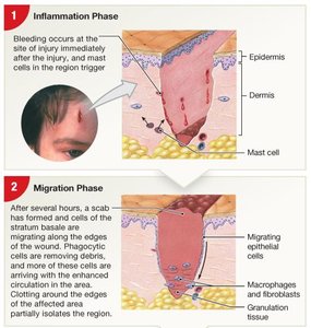
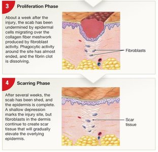
Skin repair involves four phases: inflammation, migration, proliferation, and scarring. The process depends on the type of tissue and extent of injury.
Inflammation: Bleeding and immune response.
Migration: Basal keratinocytes and fibroblasts migrate to the wound.
Proliferative: Cells divide to repair dermis and epidermis.
Scarring: Fibroblasts produce scar tissue, especially in the dermis.
Burns
Classification and Effects
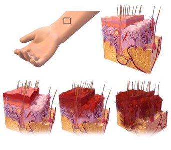
1st Degree: Damages only the epidermis; red, swollen, dry.
2nd Degree: Damages epidermis and part of dermis; blistered, painful.
3rd Degree: Damages all skin layers and accessory structures; white, blackened, or charred; may be less painful due to nerve destruction.
Effects of Aging on the Integument
Age-Related Changes
Thinner epidermis and reduced immune response increase risk of injury and infection.
Decreased vitamin D3 production affects bone and muscle health.
Reduced melanin production increases sun sensitivity.
Gland activity decreases, leading to dry, scaly skin.
Hair thins and changes color due to reduced cell division.
Wrinkles and sagging result from slower collagen replacement.
Impaired thermoregulation and slower healing due to reduced cell division and fibroblast activity.
Tattoos
Clinical Relevance
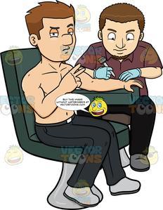
Tattoos are permanent because the ink is deposited in the dermis, which is more stable than the epidermis. Sun exposure can fade tattoos, and removal is difficult.