 Back
BackThe Integumentary System: Structure, Function, and Clinical Relevance
Study Guide - Smart Notes
 Tailored notes based on your materials, expanded with key definitions, examples, and context.
The Integumentary System
Overview and Functions
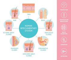
The integumentary system is the largest organ system in the human body, consisting of the skin and its accessory structures. It serves as the primary interface between the body and the external environment, providing multiple essential functions for survival and homeostasis.
Barrier Function: Protects internal tissues from mechanical damage, pathogens, and dehydration.
Immune Response: Acts as a first line of defense, containing immune cells that detect and respond to pathogens.
Vitamin D Synthesis: Skin cells synthesize vitamin D when exposed to UV light, essential for calcium metabolism.
Excretory Function: Removes waste products through sweat glands.
Sensory Role: Contains receptors for touch, pain, temperature, and pressure.
Temperature Regulation: Regulates body temperature via sweat production and blood flow adjustments.
Protection: Shields against UV radiation and chemical exposure.
Structure of the Skin
Major Layers of the Skin
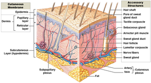
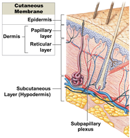
The skin is composed of three main layers, each with distinct structures and functions:
Epidermis: The outermost layer, made of closely packed epithelial cells. It provides a waterproof barrier and creates our skin tone.
Dermis: Located beneath the epidermis, this layer consists of dense irregular connective tissue, blood vessels, hair follicles, and glands.
Hypodermis (Subcutaneous Layer): Composed of loose connective and fatty tissues, it insulates the body and anchors the skin to underlying structures.
Epidermis: Structure and Layers
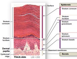
The epidermis is a keratinized stratified squamous epithelium, composed of several layers of cells. The outermost cells are dead and filled with keratin, providing a tough, protective layer.
Stratum Corneum: Outermost layer; dead, keratinized cells.
Stratum Lucidum: Present only in thick skin (palms, soles); thin, clear layer of dead cells.
Stratum Granulosum: Cells contain granules of keratohyalin; begin to die.
Stratum Spinosum: Several layers of keratinocytes; provides strength and flexibility.
Stratum Basale: Deepest layer; single row of cuboidal cells; site of cell division.
Mnemonic: "Come, Let's Get Sun Burned" (Corneum, Lucidum, Granulosum, Spinosum, Basale).
Dermis: Structure and Layers
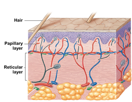
The dermis supports and nourishes the epidermis and is embedded with accessory structures. It is divided into two layers:
Papillary Layer: Composed of loose connective tissue; contains capillaries and sensory neurons; nourishes the epidermis.
Reticular Layer: Dense irregular connective tissue; contains collagen and elastin fibers, sensory receptors, and blood vessels.
Hypodermis (Subcutaneous Layer)
The hypodermis lies beneath the dermis and is highly vascularized. It serves two main roles:
Insulation: Fat cells help insulate the body and conserve heat.
Anchoring: Connects the skin to underlying muscles and bones.
Factors Influencing Skin Color
Pigments and Their Roles
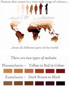
Skin color is primarily determined by the amount and type of pigments present in skin cells:
Melanin: Produced by melanocytes; two types—pheomelanin (red-yellow) and eumelanin (brown-black). Melanin protects against UV radiation.
Carotene: Yellow-orange pigment obtained from the diet; can accumulate in the epidermis.
Melanin Production and Distribution
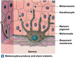
Melanocytes produce melanin and transfer it to keratinocytes via melanosomes. Melanin production increases with UV exposure, providing adaptive protection against DNA damage.
Accessory Structures of the Skin
Overview of Accessory Structures
Accessory structures are embedded in the dermis and project through the epidermis. They include:
Hair and Hair Follicles: Provide protection, insulation, and sensory input.
Exocrine Glands: Sebaceous (oil) and sudoriferous (sweat) glands; involved in lubrication, excretion, and thermoregulation.
Nails: Protect the tips of fingers and toes; composed of dead, keratinized cells.
Corpuscles: Sensory receptors for touch, pressure, and vibration.
Hair
Hair is a nonliving structure produced by hair follicles. It serves several functions:
Protection: Guards scalp and body from UV radiation and minor injury.
Cushioning and Insulation: Helps retain heat.
Sensory Reception: Detects light touch via hair root plexuses.
Hair color is determined by the type and amount of melanin produced by melanocytes in the hair matrix. The shape of the hair shaft and follicle determines whether hair is straight or curly.
Exocrine Glands
There are two main types of exocrine glands in the skin:
Sebaceous (Oil) Glands: Secrete sebum, which lubricates and protects hair and skin, and inhibits bacterial growth.
Sudoriferous (Sweat) Glands: Include eccrine (widely distributed, thermoregulation), apocrine (armpits, nipples, pubic region), mammary (milk production), and ceruminous (earwax production) glands.
Nails
Nails protect the dorsal surfaces of the fingers and toes. Growth begins at the nail root, and the nail body is composed of dead, keratinized cells. The eponychium is the cuticle, and the lunula is the white crescent at the base of the nail. Nail appearance can indicate health or disease states.
Corpuscles
Corpuscles are specialized sensory receptors in the skin:
Tactile (Meissner) Corpuscle: Detects light touch.
Lamellar (Pacinian) Corpuscle: Detects deep pressure and vibration.
Bulbous Corpuscle: Detects deep, continuous pressure.
Heat Production and Thermoregulation
Heat Production in the Body
Body temperature is regulated by balancing heat production and heat loss. Core temperature refers to the temperature of internal organs, while shell temperature refers to the surface temperature of the body.
Metabolism: The main source of heat, especially from muscles, liver, and endocrine glands.
Blood Flow: Distributes heat throughout the body.
Influencing Factors: Food intake, hormones, disease, and physical activity affect heat production.
Heat Loss Mechanisms
Most heat is lost through the skin (about 80%), with the remainder lost via the lungs and excretory products. Heat loss occurs through:
Convection: Transfer of heat by air or liquid movement.
Evaporation: Loss of heat as sweat evaporates from the skin.
Conduction: Direct transfer of heat to objects in contact with the skin.
Radiation: Emission of infrared heat rays from the body.
Thermoregulation
The hypothalamus is the control center for thermoregulation. The body can lose or conserve heat through various mechanisms:
Heat Loss: Dilation of blood vessels, movement of blood to the skin (shell), and sweating.
Heat Conservation: Constriction of blood vessels, movement of blood to the core, and shivering.
Thermoregulation in Newborns and the Elderly
Newborns: Lose heat rapidly due to a large surface area, less insulation, and limited ability to shiver. Rely on nonshivering thermogenesis (brown adipose tissue metabolism).
Elderly: Thinner skin, reduced blood supply, and decreased basal cell activity make it harder to retain or dissipate heat, increasing risk in extreme temperatures.
Summary Table: Layers of the Skin
Layer | Main Tissue Type | Key Features |
|---|---|---|
Epidermis | Keratinized stratified squamous epithelium | Protection, waterproof barrier, pigment production |
Dermis (Papillary) | Loose connective tissue | Capillaries, sensory neurons, supports epidermis |
Dermis (Reticular) | Dense irregular connective tissue | Collagen, elastin, sensory receptors, glands |
Hypodermis | Loose connective and adipose tissue | Insulation, energy storage, anchors skin |
Take Home Points
The integumentary system is the largest organ system, consisting of the skin and its accessory structures.
The skin is composed of the epidermis, dermis, and hypodermis, each with specialized functions.
Skin color is determined by melanin production from melanocytes.
Accessory structures include hair, nails, corpuscles, and exocrine glands.
Thermoregulation is essential for homeostasis and is controlled by the hypothalamus; it is impaired in newborns and the elderly.
