 Back
BackThe Integumentary System: Structure, Function, and Clinical Relevance
Study Guide - Smart Notes
 Tailored notes based on your materials, expanded with key definitions, examples, and context.
The Integumentary System
Overview and Components
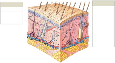
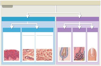
The integumentary system is the body's largest organ system, primarily responsible for protecting internal structures and maintaining homeostasis. It consists of the cutaneous membrane (skin) and accessory structures such as hair, nails, and exocrine glands.
Cutaneous Membrane: Composed of the epidermis and dermis.
Accessory Structures: Includes hair follicles, exocrine glands, and nails.
Subcutaneous Layer: Lies beneath the dermis, containing adipose tissue for insulation and energy storage.
Functions of the Integumentary System
The integumentary system serves several essential functions for the body:
Physical Protection: Shields against environmental hazards, pathogens, and mechanical injury.
Water Loss Prevention: Reduces dehydration by limiting water evaporation.
Vitamin D3 Synthesis: Initiates vitamin D3 production when exposed to sunlight.
Sensory Reception: Contains receptors for touch, pressure, pain, and temperature.
Thermoregulation: Regulates body temperature via blood vessels and sweat glands.
Excretion: Removes waste products through sweat.
Immune Response: Coordinates defense against pathogens and cancers.
Lipid Storage: Stores fats in the subcutaneous layer.
Structure of the Skin
Layers of the Skin
The skin is organized into three main layers:
Epidermis: The outermost layer, composed mainly of keratinocytes. It provides a barrier and is involved in continuous cell renewal.
Dermis: Located beneath the epidermis, it contains connective tissue, blood vessels, nerves, and accessory structures.
Subcutaneous Layer (Hypodermis): Contains adipocytes and provides insulation and energy storage.
Epidermal Layers
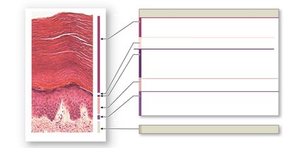
The epidermis consists of several distinct layers, each with specialized functions:
Stratum Corneum: Multiple layers of dead, flattened keratinocytes; water-resistant.
Stratum Lucidum: Present only in thick skin (palms, soles); glassy appearance.
Stratum Granulosum: Keratinocytes produce keratin; cells flatten and die.
Stratum Spinosum: Keratinocytes are bound by desmosomes; contains Langerhans cells for immunity.
Stratum Basale (Germinativum): Deepest layer; contains stem cells, melanocytes, and tactile cells.
Thin vs. Thick Skin
Thin Skin: Contains four layers of keratinocytes; found on most of the body.
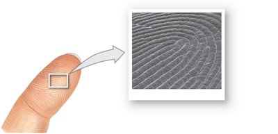
Thick Skin: Contains five layers; found on palms and soles; features prominent epidermal ridges (fingerprints).
Cells of the Epidermis
Keratinocytes
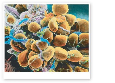
Function: Produce keratin, a protein that strengthens the skin.
Turnover: Continuously replaced; shed 30,000–40,000 cells per minute.
Melanocytes
Function: Produce melanin pigment, which protects against UV radiation.
Location: Found in the stratum basale.
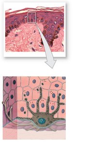
Langerhans Cells
Function: Immune defense; detect and respond to pathogens.
Tactile (Merkel) Cells
Function: Sensory reception for touch.
Dermis and Subcutaneous Layer
Dermis Structure
The dermis is divided into two layers:
Papillary Layer: Contains capillaries, nerve endings, and forms dermal papillae.
Reticular Layer: Dense connective tissue with collagen and elastic fibers; provides strength and elasticity.
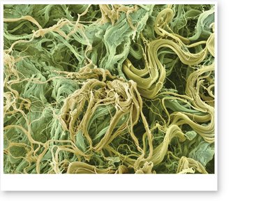
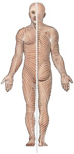
Tension (Cleavage) Lines
Definition: Patterns formed by collagen bundles and elastic fibers.
Clinical Importance: Incisions parallel to tension lines heal better; stretch marks result from rapid dermal stretching.
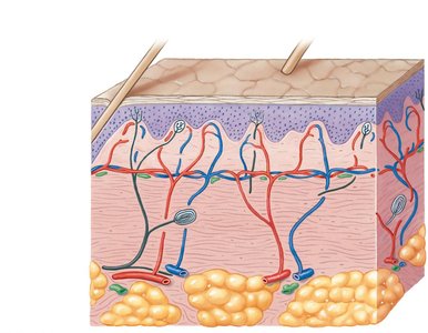
Dermal Circulation
Blood Supply: Capillary networks in the papillary and reticular layers support skin nutrition and thermoregulation.
Lymphatic Vessels: Help remove excess fluid and defend against infection.
Subcutaneous Layer (Hypodermis)
Composition: Contains adipocytes (fat cells) and connective tissue.
Function: Provides insulation, energy storage, and anchors skin to underlying tissues.
Skin Color and Pigmentation
Melanin and Skin Color
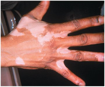
Melanin: Produced by melanocytes; determines skin color and provides UV protection.
Clinical Note: Disorders such as vitiligo result in loss of pigmentation.
Clinical Relevance
Skin Cancers
Basal Cell Carcinoma: Most common; rarely metastasizes; treated by excision.
Squamous Cell Carcinoma: Also common; low risk of metastasis.
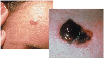
Malignant Melanoma: Highly aggressive; early detection is critical.
A: Asymmetry
B: Borders (irregular)
C: Color (varied)
D: Diameter (>6mm)
Other Clinical Conditions
Ichthyosis: Characterized by dry, scaly skin ("fish skin").
Insensible vs. Sensible Perspiration: Insensible water loss is continuous and can be life-threatening in burns; sensible perspiration increases with exercise.
Epidermal Growth Factor (EGF): Promotes skin growth and repair; used in tissue culture and grafts.
Summary Table: Layers and Functions of the Skin
Layer | Main Features | Functions |
|---|---|---|
Epidermis | Keratinocytes, melanocytes, Langerhans cells | Protection, water resistance, UV defense |
Dermis | Papillary and reticular layers, collagen, elastic fibers | Strength, elasticity, sensory reception, blood supply |
Subcutaneous Layer | Adipocytes, connective tissue | Insulation, energy storage, anchoring |
Key Equations and Concepts
Keratinocyte Turnover:
Insensible Water Loss:
