 Back
BackThe Integumentary System: Structure, Function, and Pathology - Med Term CH 16
Study Guide - Smart Notes
 Tailored notes based on your materials, expanded with key definitions, examples, and context.
The Integumentary System
Introduction
The integumentary system consists of the skin and its accessory structures, including hair, nails, and glands. It serves as the body's primary barrier against environmental hazards and plays a crucial role in sensation, thermoregulation, and metabolic functions.
Structure of the Skin
Layers of the Skin
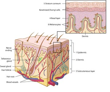
The skin is composed of three main layers, each with distinct functions and cellular compositions:
Epidermis: The outermost layer, consisting of stratified squamous epithelium. It includes the basal layer (site of cell division), stratum corneum (keratinized cells), and melanocytes (pigment-producing cells).
Dermis: The middle layer, made of dense connective tissue containing collagen fibers, blood vessels, nerve endings, and glands.
Subcutaneous Layer: The innermost layer, composed mainly of adipose tissue, providing insulation and energy storage.
Example: The basal layer continuously produces new epidermal cells, which migrate upward and become keratinized.
Accessory Structures of the Skin
Hair
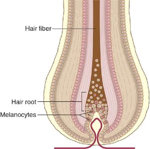
Hair is composed of keratinized cells and grows from follicles embedded in the dermis. Melanocytes at the root determine hair color. Hair serves protective and sensory functions.
Hair Follicle: The sac within which each hair grows.
Hair Root: The base of the hair, where growth occurs.
Melanocytes: Cells that produce pigment for hair color.
Nails
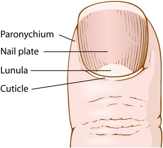
Nails are hard keratin plates covering the fingers and toes. They protect the distal phalanges and aid in fine manipulation.
Lunula: The whitish, half-moon-shaped area at the base of the nail.
Cuticle: The band of epidermis at the base and sides of the nail plate.
Paronychium: The soft tissue surrounding the nail border.
Glands
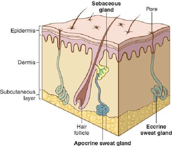
The skin contains two main types of glands:
Sebaceous Glands: Secrete oily sebum into hair follicles to lubricate skin and hair.
Sweat Glands: Include eccrine (most numerous, produce watery sweat for cooling) and apocrine (located in axilla and genital areas, produce thicker secretions).
Functions of the Skin
Protective and Regulatory Functions
The skin acts as a protective membrane, regulates body temperature, and serves as a receptor for sensations such as pain, temperature, pressure, and touch. It also produces sweat and sebum, which help cool and lubricate the skin.
Thermoregulation: Sweat glands produce a watery secretion that evaporates to cool the body.
Sensation: Nerve endings in the skin detect environmental stimuli.
Barrier: Prevents entry of pathogens and loss of water.
Medical Terminology Related to Dermatology
Combining Forms and Colors
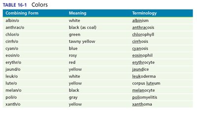
Medical terms often use combining forms to describe colors and conditions of the skin. Understanding these forms aids in interpreting medical reports.
Combining Form | Meaning | Terminology |
|---|---|---|
albin/o | white | albinism |
melan/o | black | melanocyte |
xanth/o | yellow | xanthoma |
erythr/o | red | erythrocyte |
cyan/o | blue | cyanosis |
leuk/o | white | leukoderma |
chlor/o | green | chlorophyll |
anthrac/o | black (as coal) | anthracosis |
jaund/o | yellow | jaundice |
Cutaneous Lesions
Types of Lesions
Lesions are abnormal changes in the skin's structure or appearance. Common types include:
Crust (scab): Dried serum and cellular debris.
Cyst: Closed sac containing fluid or semisolid material.
Erosion: Loss of superficial epidermis.
Fissure: Deep groove or crack.
Macule: Flat, colored spot.
Nodule: Solid, raised lesion.
Papule: Small, raised bump.
Pustule: Elevated lesion containing pus.
Ulcer: Open sore with loss of tissue.
Vesicle: Small, fluid-filled blister.
Wheal: Raised, red area due to allergic reaction.
Signs and Symptoms of Skin Disorders
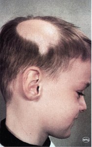
Alopecia
Alopecia is the absence of hair where it normally grows, often resulting from autoimmune or genetic factors.
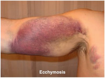
Ecchymosis
Ecchymosis refers to blue-purple marks on the skin, commonly known as bruises, caused by bleeding under the skin.
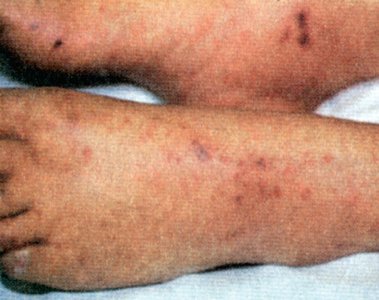
Petechia
Petechia are small, pinpoint hemorrhages resulting from capillary bleeding.
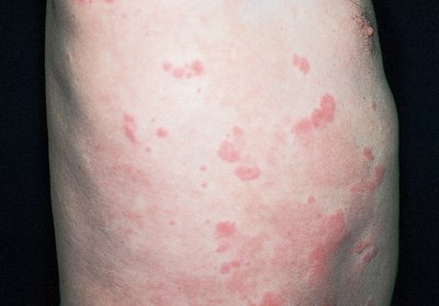
Urticaria
Urticaria (hives) is an acute allergic reaction characterized by red, round wheals on the skin.
Abnormal Skin Conditions
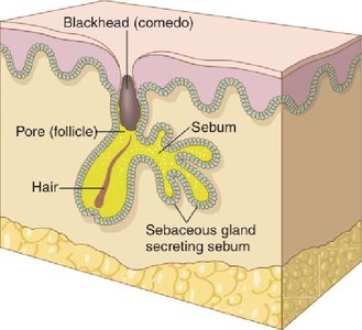
Acne
Acne is a papular and pustular eruption of the skin with increased production of sebum, often resulting in blackheads and inflammation.
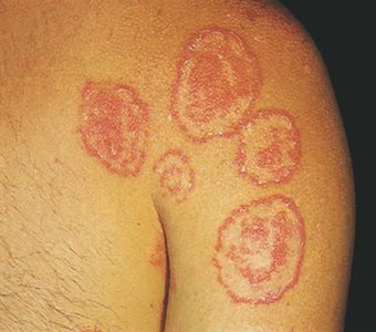
Tinea
Tinea is a fungal infection of the skin, commonly known as ringworm. It presents as circular, red, scaly patches.
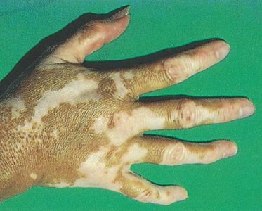
Vitiligo
Vitiligo is the loss of pigment in areas of the skin, resulting in white patches.
Skin Neoplasms
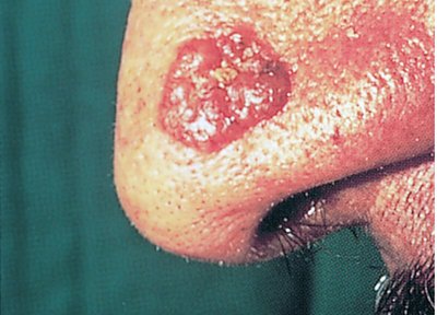
Basal Cell Carcinoma
Basal cell carcinoma is a malignant tumor of the basal cell layer of the epidermis. It is the most common type of skin cancer and typically appears as a raised, pearly lesion.
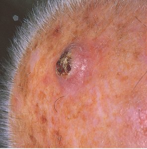
Squamous Cell Carcinoma
Squamous cell carcinoma is a malignant tumor of the squamous epithelial cells of the epidermis. It often presents as a scaly, red patch or nodule.
Laboratory Tests and Clinical Procedures
Laboratory Tests
Bacterial Analyses: Examination of pus or fluid samples to detect microorganisms.
Fungal Tests: Skin scrapings are cultured and examined microscopically after treatment with potassium hydroxide (KOH).
Clinical Procedures
Cryosurgery: Destruction of tissue using subzero temperatures (liquid nitrogen).
Curettage: Scraping of lesions with a sharp curet.
Electrodesiccation: Destruction of tissue by burning with an electric spark.
Mohs Surgery: Removal of thin layers of growth for microscopic examination, especially for basal and squamous cell carcinomas.
Skin Biopsy: Removal of skin tissue (punch or shave) for pathological examination.
Skin Test: Testing the body's reaction to allergens (scratch or patch tests).
Abbreviations in Dermatology
Abbreviation | Meaning |
|---|---|
ABCDE | Asymmetry, Border, Color, Diameter, Evolution (characteristics of melanoma) |
Bx | Biopsy |
Derm. | Dermatology |
DLE | Discoid lupus erythematosus |
PPD | Purified protein derivative |
PUVA | Psoralen-ultraviolet A light therapy |
SLE | Systemic lupus erythematosus |
SC | Subcutaneous |
Suffixes Commonly Used in Dermatology
Suffix | Meaning |
|---|---|
-algia | Pain |
-derma | Skin |
-esis | Condition |
-lysis | Breakdown; separation; destruction; loosening |
-ose | Full of; pertaining to; sugar |
-osis | Condition, usually abnormal |
-ous | Pertaining to |
-plakia | Plaque |
-plasty | Surgical repair |
-rrhea | Flow; discharge |
Summary
The integumentary system is essential for protection, sensation, and regulation. Understanding its structure, functions, and common pathologies is fundamental for students in anatomy and physiology courses.
