 Back
BackThe Integumentary System: Structure, Function, and Clinical Relevance
Study Guide - Smart Notes
 Tailored notes based on your materials, expanded with key definitions, examples, and context.
The Integumentary System
Overview and Importance
The integumentary system is the body's primary protective barrier, consisting of the skin and its derivatives. It plays crucial roles in protection, sensation, thermoregulation, metabolic functions, and excretion. Understanding this system is essential for evaluating and treating skin injuries, such as burns.
Structure of the Skin
Main Regions of the Skin
Epidermis: The superficial region composed of keratinized stratified squamous epithelium.
Dermis: The deeper region, primarily made of fibrous dense irregular connective tissue.
Hypodermis (Superficial Fascia): Subcutaneous layer deep to the skin, mostly adipose tissue. Not part of the skin but shares some functions, such as insulation, protection, and anchoring the skin to underlying structures.
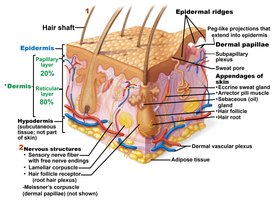
Skin Structure and Appendages
Hair: Sensory touch receptors, protection from physical trauma, heat loss, and sunlight.
Nails: Protective coverings for fingers and toes.
Sweat and Sebaceous Glands: Involved in thermoregulation and lubrication of skin and hair.
Epidermis: Layers and Cell Types
Layers of the Epidermis
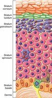
The epidermis consists of four or five distinct layers (from superficial to deep):
Stratum corneum: 20–30 rows of dead, flat, keratinized membranous sacs.
Stratum lucidum: Thin, translucent band present only in thick skin.
Stratum granulosum: One to five cell layers where keratinization begins.
Stratum spinosum: Several layers thick, containing intermediate filaments and desmosomes for strength.
Stratum basale: Deepest layer, single row of stem cells, actively mitotic, contains melanocytes.
Cell Types in the Epidermis
Keratinocytes: Produce keratin, most abundant, arise from stratum basale, connected by desmosomes.
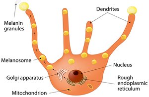
Melanocytes: Produce melanin, protect against UV damage, found in stratum basale.
Dendritic (Langerhans) cells: Phagocytes and key activators of the immune system.
Tactile epithelial (Merkel) cells: Sensory touch receptors at the dermis-epidermal junction.
Dermis: Layers and Features
Papillary Layer
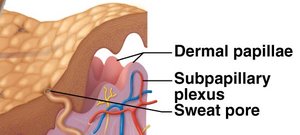
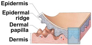
The papillary layer is composed of areolar connective tissue with collagen and elastic fibers. It contains dermal papillae, which are peg-like projections that extend into the epidermis, providing nutrients and oxygen to lower layers and housing sensory receptors.
Reticular Layer
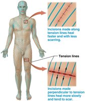
The reticular layer makes up about 80% of the dermis and consists of dense irregular connective tissue. Collagen fibers provide strength and resiliency, while elastic fibers allow stretch and recoil. Gaps between collagen bundles form cleavage (tension) lines, important for surgical incisions.
Skin Markings
Friction ridges: Enhance gripping ability and contribute to fingerprints.
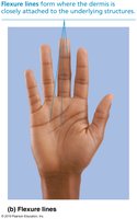
Flexure lines: Dermal folds at or near joints, visible on hands and feet.
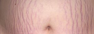
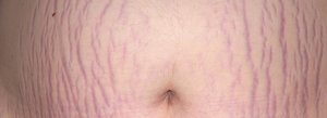
Striae: Stretch marks from dermal tears.
Blisters: Fluid-filled pockets separating epidermal and dermal layers.
Skin Color
Pigments Determining Skin Color
Melanin: Only pigment made in skin, ranges from reddish-yellow to brownish-black. Produced by melanocytes, packed into melanosomes, and transferred to keratinocytes.
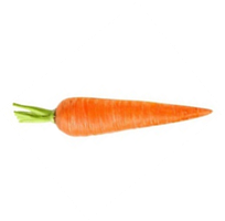
Carotene: Yellow to orange pigment from plant products, accumulates in stratum corneum and hypodermis.
Hemoglobin: Iron-containing red pigment in red blood cells, gives pinkish hue to fair skin.
Clinical Significance of Skin Color
Cyanosis: Bluish-gray tint due to poorly oxygenated hemoglobin.
Pallor: Pale color from emotional stress, low blood pressure, or anemia.
Erythema: Reddened skin from embarrassment, exercise, inflammation, allergy, or fever.
Jaundice: Yellow discoloration from liver disorder and bilirubin accumulation.
Bruises: Red/purple/green/yellow marks from clotted blood beneath skin.
Dark patches: May signal endocrine disorders such as diabetes mellitus.
Hair and Its Functions
Structure and Function of Hair
Hair: Composed of dead keratinized cells of hard keratin, more durable than skin keratin.
Functions: Sensory touch receptors, protection from physical trauma, heat loss, and sunlight.
Hair Color and Types
Hair pigments: Melanins (yellow, rust, brown, black) and pheomelanin (red hair).
Gray/white hair: Decreased melanin production, air bubbles replace melanin in shaft.
Hair types: Vellus (fine, pale) and terminal (coarse, long).
Hair Growth and Disorders
Growth cycles: Active and resting phases, nutrition and hormones affect growth.
Alopecia: Thinning of hair with age, follicles have limited cycles.
True baldness: Male pattern baldness, genetically determined, sex-linked.
Hirsutism: Excessive hair growth in females, often due to androgen excess.
Sweat and Sebaceous Glands
Sweat Glands
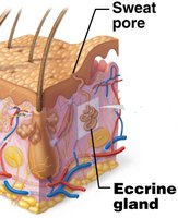
Eccrine sweat glands: Most numerous, abundant on palms, soles, and forehead. Function in thermoregulation, secrete hypotonic sweat.
Apocrine sweat glands: Confined to axillary and anogenital areas, secrete sweat plus fatty substances and proteins. Begin functioning at puberty, may act as sexual scent glands.
Modified apocrine glands: Ceruminous (earwax) and mammary (milk) glands.
Sebaceous (Oil) Glands
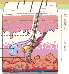
Distribution: Widely distributed except in thick skin of palms and soles.
Function: Secrete sebum, an oily, acidic holocrine secretion that softens, lubricates, waterproofs skin, and has bactericidal action.
Functions of the Integumentary System
Major Functions
Protection: Biological, chemical, and physical barriers.
Temperature regulation: Sweat glands and dermal blood vessels regulate heat loss.
Cutaneous sensation: Sensory receptors respond to pain, pressure, touch, and temperature.
Metabolic functions: Synthesis of vitamin D precursor.
Blood reservoir: Dermal vascular supply holds 5% of blood volume.
Excretion: Loss of nitrogenous wastes in sweat (ammonia, urea, uric acid).
Skin Cancer
Major Types
Basal cell carcinoma: Most common, least malignant, arises from stratum basale.
Squamous cell carcinoma: Second most common, arises from keratinocytes of stratum spinosum.
Melanoma: Cancer of melanocytes, least common but most dangerous, highly metastatic.
ABCDE Rule for Melanoma Detection
A: Asymmetry
B: Border irregularity
C: Color variation
D: Diameter > 6 mm
E: Evolving (changing in size, shape, color)
Burns
Classification and Severity
First-degree burns: Affect only the epidermis.
Second-degree burns: Affect epidermis and part of dermis.
Third-degree burns: Full thickness, affect epidermis, dermis, and sometimes deeper tissues.
Rule of Nines
The percentage of body surface burned is estimated using the Rule of Nines, dividing the body into 11 areas, each accounting for 9% of total body area, plus 1% for the genital area.
Area | % of Body Surface |
|---|---|
Head and Neck | 9% |
Each Arm | 9% |
Each Leg | 18% |
Anterior Trunk | 18% |
Posterior Trunk | 18% |
Perineum | 1% |
Burns are considered critical if over 25% of the body has second-degree burns, over 10% has third-degree burns, or third-degree burns are present on hands, feet, or face.
Summary Table: Layers of the Skin
Layer | Location | Main Features |
|---|---|---|
Epidermis | Superficial | Keratinized stratified squamous epithelium |
Dermis | Deep to epidermis | Fibrous dense irregular connective tissue |
Hypodermis | Deep to dermis | Adipose tissue, anchors skin, not part of skin |
Additional info: Academic context was added to clarify the functions, clinical relevance, and structural details of the integumentary system, as well as to provide self-contained explanations suitable for exam preparation.
