 Back
BackThe Integumentary System: Structure, Function, and Clinical Correlates
Study Guide - Smart Notes
 Tailored notes based on your materials, expanded with key definitions, examples, and context.
The Integumentary System
Overview and Functions
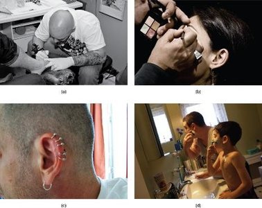
The integumentary system consists of the skin and its accessory structures, including hair, nails, and glands. It is the largest organ system in the human body, accounting for approximately 16% of body weight and covering 1.5 to 2 m2. The skin serves as a protective barrier, provides immune defense, synthesizes hormones, maintains fluid balance, and regulates body temperature.
Protection: Shields the body from mechanical damage, pathogens, UV radiation, and dehydration.
Sensory Input: Contains nerve endings and receptors for touch, pain, temperature, and pressure.
Thermoregulation: Modulates heat loss through vasodilation, vasoconstriction, sweating, and insulation.
Vitamin D Synthesis: Initiates the production of vitamin D upon exposure to UV light.
Structure of the Skin
Major Layers
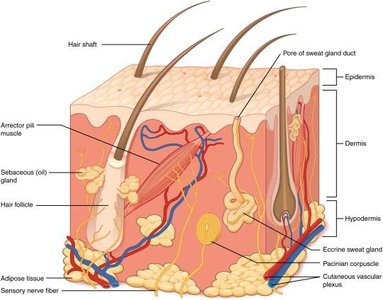
The skin is composed of three primary layers: the epidermis, dermis, and hypodermis. Each layer has distinct structural and functional characteristics.
Epidermis: Outermost layer, composed of keratinized, stratified squamous epithelium. It is avascular and primarily consists of keratinocytes.
Dermis: Middle layer, made of dense irregular connective tissue. It houses blood vessels, nerves, hair follicles, and glands.
Hypodermis (Subcutaneous Layer): Deepest layer, composed mainly of adipose and loose areolar connective tissue. Provides insulation, cushioning, and energy storage.
Microscopic Structure: Thin vs. Thick Skin
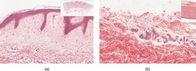
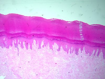
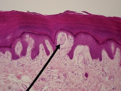
Thin skin covers most of the body and has four layers in the epidermis, while thick skin (palms and soles) has five layers and a much thicker epithelial layer.
The Epidermis
Layers of the Epidermis
The epidermis is organized into distinct layers (strata), each with specialized cells and functions. From deep to superficial, the layers are:
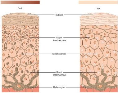
Stratum Basale: Single row of cuboidal cells undergoing mitosis. Contains melanocytes (produce melanin for UV protection) and Merkel cells (detect light touch).
Stratum Spinosum: 8–10 layers of cells connected by desmosomes, appearing spiny. Contains Langerhans (dendritic) cells for immune defense.
Stratum Granulosum: 3–5 layers of flattened cells producing keratin and keratohyalin. Cells lose nuclei and organelles as they move upward.
Stratum Lucidum: Present only in thick skin. Translucent layer of dead cells filled with eleidin protein.
Stratum Corneum: 15–30 layers of dead, keratinized cells that provide a tough, protective barrier.
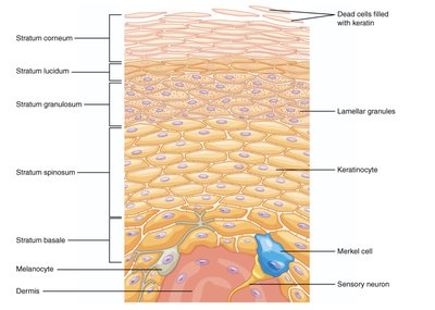
Specialized Cells of the Epidermis
Keratinocytes: Main cell type, produce keratin for strength and waterproofing.
Melanocytes: Produce melanin pigment, protecting DNA from UV damage and determining skin color.
Merkel Cells: Sensory receptors for light touch, most abundant in hands and feet.
Langerhans Cells: Immune cells that capture pathogens and debris.
The Dermis
Layers of the Dermis
The dermis is divided into two layers:
Papillary Dermis: Superficial layer of loose areolar connective tissue. Contains dermal papillae (fingerlike projections), capillaries, free nerve endings, and Meissner's corpuscles (touch receptors). Responsible for fingerprints.
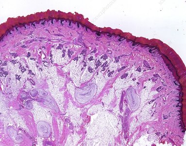
Reticular Dermis: Deeper layer of dense irregular connective tissue. Contains collagen fibers for strength and elasticity, hair follicles, sweat glands, and a rich nerve and vascular supply.
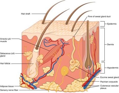
Sensory Nerve Receptors in the Dermis
Free Nerve Endings: Detect pain and temperature.
Meissner's (Tactile) Corpuscles: Detect discriminative touch, located in dermal papillae.
Ruffini's (Bulbous) Corpuscles: Detect stretch and slippage.
Pacinian (Lamellar) Corpuscles: Detect deep pressure and vibration, located deep in the dermis.
Root Hair Plexus: Detects movement of hair.
The Hypodermis
Structure and Function
The hypodermis, or subcutaneous layer, lies beneath the dermis. It is primarily composed of adipose tissue and loose areolar connective tissue. This layer provides thermal insulation, cushioning, and serves as an energy reservoir.
Skin Pigmentation
Determinants of Skin Color
Skin color is primarily determined by the amount and type of melanin produced by melanocytes in the stratum basale. Other pigments and physiological factors also contribute to skin coloration.
Melanin: Protects DNA from UV radiation. More melanin results in darker skin; less melanin results in lighter skin.
Carotene: Contributes a yellowish tint.
Pheomelanin: Responsible for red hair and ruddy skin tones.
Hemoglobin: Oxygenation state affects skin color (e.g., cyanosis, flushing).
Clinical Variants and Disorders of Pigmentation
Freckles: Localized accumulations of melanin.
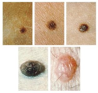
Nevi (Moles): Concentrated areas of melanocytes; should be monitored for changes.
Poliosis: White patches of hair lacking melanin.
Albinism: Genetic lack of melanin production.
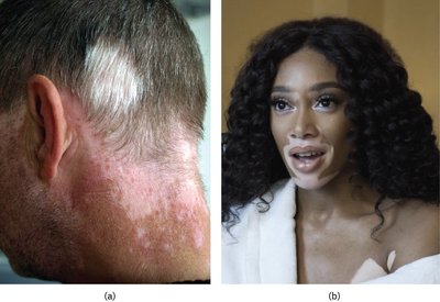
Vitiligo: Autoimmune destruction of melanocytes, causing depigmented patches.
Melanoma: Malignant tumor of melanocytes.
Other Color Variants
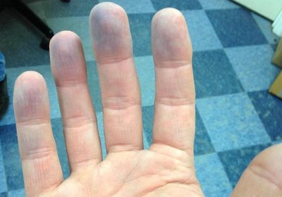
Cyanosis: Bluish tint due to low oxygenation, most visible in lips and fingertips.
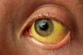
Jaundice: Yellowing of skin and eyes due to bilirubin accumulation from liver dysfunction.
Pallor: Pale skin from reduced blood flow or anemia.
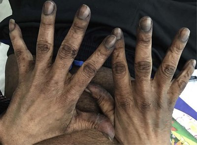
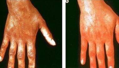
Addison’s Disease: Adrenal insufficiency causing bronzing hyperpigmentation.
Accessory Structures of the Skin
Hair
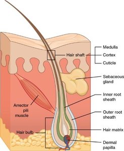
Hair is produced by hair follicles originating in the epidermis and extending into the dermis. Hair serves protective, sensory, and thermoregulatory functions.
Hair Shaft: Visible, non-anchored portion.
Hair Root and Bulb: Anchored in the dermis; contains the hair matrix (site of cell division).
Medulla, Cortex, Cuticle: Structural layers of the hair shaft.
Arrector Pili Muscle: Smooth muscle that causes hair to stand (goosebumps).
Nails
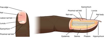
Nails are protective coverings on the dorsal surface of fingers and toes, composed of hard keratin. They aid in grasping objects and protecting the distal phalanges.
Glands
Eccrine Sweat Glands: Widely distributed, secrete watery sweat for thermoregulation via merocrine secretion.
Apocrine Sweat Glands: Located in axilla and groin, secrete a thicker fluid into hair follicles; function in scent communication.
Sebaceous Glands: Secrete sebum (lipid-rich) into hair follicles for lubrication and waterproofing; become active at puberty.
Functions of the Integument
Thermoregulation
The skin regulates body temperature through several mechanisms:
Heat Release: Vasodilation, sweating (evaporation), radiation, convection, and conduction.
Heat Conservation: Vasoconstriction reduces blood flow to the skin, minimizing heat loss.
Vitamin D Synthesis
Upon exposure to UV light, cholesterol in the skin is converted to cholecalciferol (vitamin D3 precursor), which is then processed by the liver and kidneys to form active vitamin D3 (calcitriol). This hormone is essential for calcium absorption and bone health.
Deficiency: Leads to rickets in children and osteomalacia in adults.
Risk Factors: Darker skin, limited sun exposure, and poor diet increase risk of deficiency.
Disorders and Diseases of the Skin
Skin Cancers
Basal Cell Carcinoma: Originates in the stratum basale; most common and least dangerous.
Squamous Cell Carcinoma: Originates in the stratum spinosum; can metastasize if untreated.
Melanoma: Originates in melanocytes; highly malignant and aggressive.
Other Disorders
Eczema: Chronic inflammatory skin condition with red, flaky rash.
Acne: Overproduction of sebum leads to clogged pores and inflammation.
Injuries
Contusion (Bruise): Bleeding under the skin due to damaged blood vessels.
Petechiae: Small red dots from broken capillaries.
Laceration: Cut through the skin; healing depends on alignment with lines of cleavage (collagen patterns).
Burns: Classified by depth (first to fourth degree). The extent is estimated by the "rule of nines" or "rule of palm." Full-thickness burns (third and fourth degree) require medical intervention for healing.
