 Back
BackThe Integumentary System: Structure, Function, and Clinical Relevance
Study Guide - Smart Notes
 Tailored notes based on your materials, expanded with key definitions, examples, and context.
The Integumentary System
Overview and Components
The integumentary system is the body's largest organ system, serving as a protective barrier and playing vital roles in physiology and health. It consists of the skin (cutaneous membrane) and its derivatives: hair, nails, sweat glands, and sebaceous glands. The scientific study of the skin is called dermatology.
Skin (Integument): Covers the body, accounts for 7-8% of body weight, and ranges from 1.5-2.0 m2 in area and 1.5-4.0 mm in thickness.
Derivatives: Hair, nails, sweat glands, sebaceous glands.
Functions: Protection, sensation, thermoregulation, metabolic functions, and more.
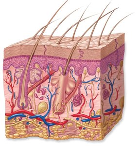
Structure of the Integument
Layers of the Skin
The skin is composed of three main layers, each with distinct structures and functions:
Epidermis: Superficial, avascular, composed of keratinized stratified squamous epithelium.
Dermis: Deeper, vascular, primarily dense irregular connective tissue.
Hypodermis (Subcutaneous Layer): Not part of the integument, but closely associated; consists of areolar and adipose tissue, providing insulation and shock absorption.
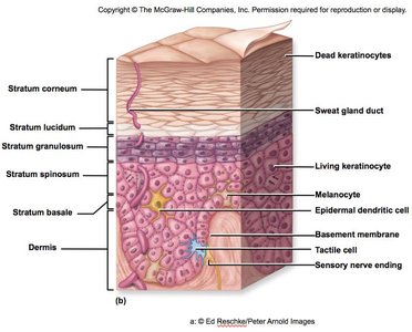
Epidermal Strata
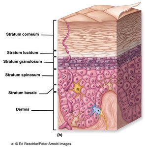
The epidermis is organized into five distinct layers (strata) from deep to superficial:
Stratum basale
Stratum spinosum
Stratum granulosum
Stratum lucidum (only in thick skin)
Stratum corneum
The first three layers contain living keratinocytes, while the outer two consist of dead, keratinized cells.
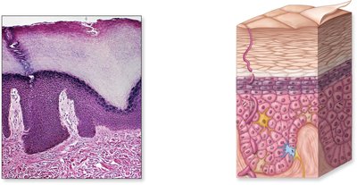
Stratum Basale
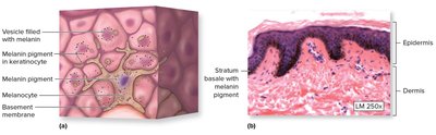
The deepest epidermal layer, also called the basal layer, is a single row of cuboidal to low columnar cells attached to the basement membrane. It contains three main cell types:
Keratinocytes: Most abundant; produce keratin for strength and waterproofing.
Melanocytes: Produce melanin pigment, protecting DNA from UV radiation.
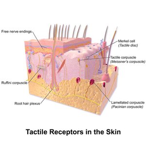
Tactile (Merkel) cells: Sensory cells sensitive to touch.
Dendritic (Langerhans) cells are also present, functioning as antigen-presenting cells for immune defense.
Stratum Spinosum
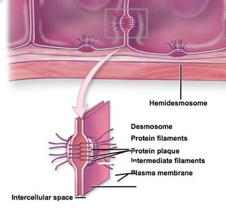
This layer consists of several layers of polygonal keratinocytes, some of which are still dividing. Cells are connected by desmosomes, providing resistance to tension and pulling. Epidermal dendritic cells are also found here.
Stratum Granulosum
Composed of 3-5 layers of keratinocytes, this is the first layer where keratinization occurs. Keratinocytes fill with keratin, lose their nuclei and organelles, and die, but remain structurally intact.
Stratum Lucidum
This thin, translucent layer is found only in thick skin (palms, soles). It consists of 2-3 layers of keratinocytes filled with eleidin, an intermediate in keratin maturation.
Stratum Corneum
The outermost layer, consisting of 20-30 layers of dead, tightly packed, anucleate keratinized cells. It forms a tough, protective barrier and is unsuitable for microbial growth due to antimicrobial peptides and glandular secretions.
Keratinocyte Migration and Skin Renewal
Keratinocytes originate in the stratum basale, migrate upward through the strata over two weeks, undergo apoptosis, and are eventually shed as dandruff or dander. Humans can shed approximately 50,000 skin cells per minute.
Thick vs. Thin Skin
Feature | Thick Skin | Thin Skin |
|---|---|---|
Location | Palms, soles, fingers, toes | Most of the body |
Strata | All five layers | Lacks stratum lucidum |
Glands | Sweat glands only | Sweat and sebaceous glands |
Hair follicles | Absent | Present |
Thickness | 0.4–0.6 mm | 0.075–0.150 mm |
Skin Color and Markings
Melanin: Brown-black to reddish-yellow pigment produced by melanocytes; protects against UV damage.
Carotene: Yellow-orange pigment from diet; accumulates in subcutaneous fat and stratum corneum; precursor to vitamin A.
Hemoglobin: Oxygen-binding protein in red blood cells; gives skin a reddish tint, especially in fair-skinned individuals.
Common skin markings include:
Nevus (mole): Benign overgrowth of melanocytes.
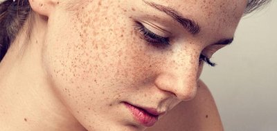
Freckles: Localized areas of increased melanocyte activity.
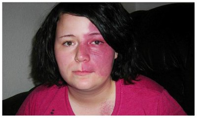
Hemangiomas: Benign blood vessel tumors (capillary or cavernous types).
Friction ridges: Unique patterns on fingers, palms, soles, and toes (fingerprints).
Clinical Aspects of Skin Color
Cyanosis: Blue skin due to low oxygenation.
Pallor: Pale color from anemia, low blood pressure, or emotional states.
Erythema: Redness from fever, inflammation, or allergy.
Jaundice: Yellow cast from liver disorders (bilirubin accumulation).
Dermis
Structure and Layers
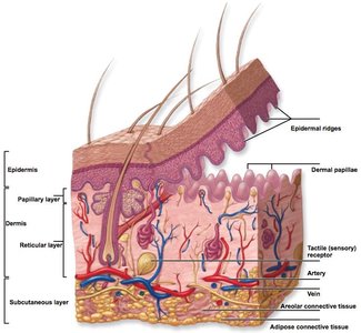
The dermis lies deep to the epidermis and is composed of strong, flexible connective tissue. It contains collagen, elastic, and reticular fibers, blood and lymphatic vessels, nerves, glands, and hair follicles. The dermis is divided into two layers:
Papillary layer: Superficial, areolar connective tissue, forms dermal papillae that interlock with epidermal ridges.
Reticular layer: Deeper, dense irregular connective tissue, provides strength and elasticity.
Lines of Cleavage and Skin Characteristics
Lines of cleavage (tension lines): Orientation of collagen and elastic fibers; important for surgical incisions (parallel incisions heal better).
Stretch marks (striae): Result from overstretched skin tearing collagen fibers.
Wrinkles: Caused by breakdown of collagen and elastic fibers, aging, and UV exposure.
Innervation and Blood Supply
Nerve fibers: Sensory (detect stimuli) and motor (control glands and blood flow).
Blood vessels: Supply nutrients, regulate temperature and blood pressure via vasoconstriction and vasodilation.
Subcutaneous Layer (Hypodermis)
This layer is not part of the integument but supports it. It consists of areolar and adipose tissue, providing padding, energy storage, and insulation. Thickness and distribution vary by sex.
Integumentary Derivatives
Nails
Nails are scalelike modifications of the stratum corneum, protecting distal digits and aiding in grasping. Key structures include the nail plate, nail bed, nail matrix (growth area), lunula, eponychium (cuticle), and hyponychium.
Hair
Types: Lanugo (fetal), vellus (fine, body hair), terminal (coarse, scalp, eyebrows, pubic regions).
Structure: Shaft (above skin), root (below skin), bulb (base, contains matrix for growth).
Follicle: Tube surrounding root, with connective and epithelial sheaths.
Arrector pili muscle: Smooth muscle causing hair to stand (goosebumps).
Functions: Protection, heat retention, sensory reception, visual identification, pheromone dispersal.
Growth: Cycles of growth and dormancy; hair loss can be normal or pathological (alopecia, male pattern baldness).
Exocrine Glands of the Skin
Merocrine (eccrine) sweat glands: Widely distributed, secrete watery sweat for thermoregulation and excretion.
Apocrine sweat glands: Found in axillae, nipples, pubic/anal regions; secrete viscous, odor-producing fluid into hair follicles.
Ceruminous glands: Modified apocrine glands in the ear canal, produce cerumen (earwax).
Mammary glands: Modified apocrine glands in breasts, produce milk during lactation.
Sebaceous glands: Produce sebum (oily secretion), lubricates skin/hair, bactericidal, holocrine secretion, activated by hormones.
Functions of the Integument
Epidermal Functions
Protection: Physical, chemical, and biological barrier.
Prevention of water loss: Keratin and lipids limit water loss; dehydration risk with burns.
Metabolic regulation: Synthesis of vitamin D3 (cholecalciferol) upon UV exposure, converted to calcitriol (active form) in liver and kidneys, essential for calcium and phosphate absorption. Equation for vitamin D activation:
Secretion and absorption: Sweat (excretion of wastes), sebum (lubrication), selective absorption (e.g., transdermal drugs).
Immune function: Dendritic cells initiate immune responses against pathogens and cancer cells.
Dermal Functions
Temperature regulation: Blood vessel dilation/constriction and sweat gland activity regulate heat loss.
Sensory reception: Numerous receptors detect touch, pressure, pain, and temperature.
Repair, Regeneration, and Aging
Wound Healing
Regeneration: Replacement of damaged cells with the same type, restoring function.
Fibrosis: Replacement with scar tissue (collagen), function not fully restored.
Aging and Clinical Considerations
Reduced stem cell activity, thinner skin, slower repair, decreased collagen and elasticity, increased wrinkles.
Decreased immune response, hair thinning/loss.
UV radiation accelerates aging and increases skin cancer risk.
Skin cancer is the most common cancer, especially in fair-skinned individuals; prevention includes sunscreen and regular exams.
