 Back
BackThe Integumentary System: Structure, Function, and Accessory Organs
Study Guide - Smart Notes
 Tailored notes based on your materials, expanded with key definitions, examples, and context.
The Integumentary System
Overview
The integumentary system is the body's largest organ system, consisting of the skin and its accessory structures. It serves as a protective barrier, regulates temperature, and provides sensory information. The skin is composed of three main layers: the epidermis, dermis, and hypodermis, each with specialized cells and functions.
The Epidermis
Cells of the Epidermis
The epidermis is the outermost layer of the skin, composed primarily of epithelial tissue and several specialized cell types:
Keratinocytes: The most abundant cells, producing keratin, a fibrous protein that protects, waterproofs, and strengthens the skin.
Melanocytes: Located in the basal layer, these cells synthesize melanin, a pigment that protects against ultraviolet (UV) radiation.
Tactile epithelial cells (Merkel cells): Found at the epidermal-dermal junction, these cells are associated with sensory nerve endings and function as touch receptors.
Dendritic (Langerhans) cells: Immune cells originating in the bone marrow that migrate to the epidermis, providing defense against pathogens.
Layers of the Epidermis
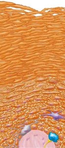
The epidermis is organized into distinct layers, each with specific functions:
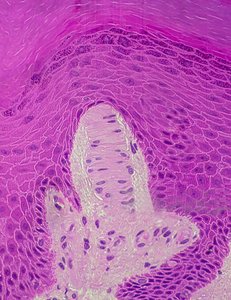
Stratum Basale: The deepest, single-cell layer where new keratinocytes are produced via mitosis. Contains melanocytes and Merkel cells. Epidermal pegs are downward projections that interlock with the dermis for structural support.
Stratum Spinosum: Several layers thick, this region is where keratinocyte maturation occurs.
Stratum Granulosum: Site of keratinization; keratohyalin granules fill with keratin, and cells begin to die.
Stratum Lucidum: A thin, clear layer only visible in thick skin (e.g., palms, soles), providing additional protection.
Stratum Corneum: The outermost, thickest layer (20–30 cell layers) composed of dead, keratinized cells that provide waterproofing and protection. Cells in this layer are shed as dandruff or dander.
The Dermis and Hypodermis
Structure and Layers of the Dermis
The dermis is the thick, supportive layer beneath the epidermis, composed mainly of connective tissue and housing most accessory structures.
Papillary Dermis: Made of areolar connective tissue, highly vascularized, and contains pain and touch receptors. Dermal papillae are projections that interlock with the epidermis, increasing surface area for exchange and support.
Reticular Dermis: Composed of dense irregular connective tissue, containing collagen and elastic fibers. This layer forms cleavage (tension) lines and flexure lines (deep creases at joints).
Hypodermis (Subcutaneous Layer): Lies beneath the dermis, consisting of adipose and areolar connective tissue. Functions include fat storage, insulation, shock absorption, and anchoring the skin to underlying tissues.
Special Features of the Dermis
Friction ridges: Form fingerprints; develop during fetal growth.
Cleavage lines: Natural patterns of tension in the skin, important for surgical incisions.
Flexure lines: Deep skin folds at joints, allowing movement.
Accessory Integumentary Structures
Hair and Hair Follicles
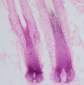
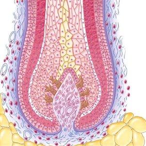
Hair is a filamentous structure composed of dead, keratinized cells. The hair follicle is a complex organ that anchors each hair into the skin.
Hair root: The portion embedded in the skin, consisting of the medulla (core), cortex (middle layer), and cuticle (outer layer).
Hair shaft: The visible part above the skin surface.
Hair follicle wall: Composed of internal and external epithelial root sheaths and a connective tissue root sheath.
Hair papilla: A structure at the base of the follicle containing blood vessels that nourish the growing hair.
Arrector pili muscle: Smooth muscle attached to the follicle; contraction causes "goosebumps."
Cutaneous Glands
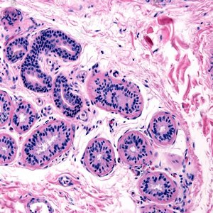
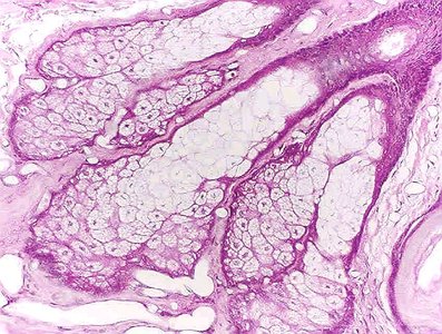
Sebaceous (Oil) Glands: Associated with hair follicles, these glands secrete sebum, an oily substance that lubricates and waterproofs the skin and hair. Function: Prevents drying and inhibits bacterial growth.
Sweat (Sudoriferous) Glands: Two main types:
Eccrine glands: Widely distributed, secrete watery sweat for thermoregulation via merocrine secretion.
Apocrine glands: Found in axillary and genital areas, secrete a thicker, milky sweat via apocrine secretion; become active at puberty.
Cutaneous Sensory Receptors
Meissner’s (Tactile) Corpuscles: Located in the papillary dermis, these receptors detect light touch and texture.
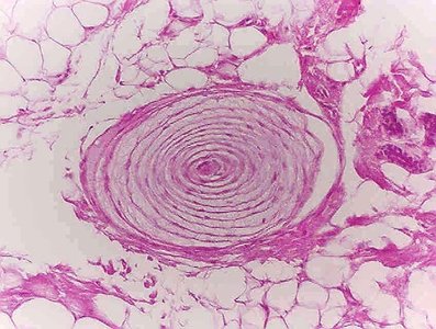
Lamellar (Pacinian) Corpuscles: Found deeper in the dermis or hypodermis, these receptors sense deep pressure and vibration.
Summary Table: Key Structures of the Integumentary System
Structure | Location | Function |
|---|---|---|
Keratinocyte | Epidermis | Produces keratin for protection and waterproofing |
Melanocyte | Stratum basale | Produces melanin for UV protection |
Merkel cell | Epidermal-dermal junction | Sensory receptor for touch |
Dendritic cell | Epidermis | Immune defense |
Sebaceous gland | Dermis, near hair follicles | Secretes sebum for lubrication |
Eccrine gland | Dermis, widely distributed | Thermoregulation via sweat |
Apocrine gland | Axillary/genital regions | Secretes milky sweat |
Meissner’s corpuscle | Papillary dermis | Detects light touch |
Lamellar corpuscle | Deep dermis/hypodermis | Detects deep pressure/vibration |
Additional Info
Goosebumps: Caused by contraction of arrector pili muscles, pulling hair upright in response to cold or emotional stimuli.
Freckles: Small, flat spots of concentrated melanin, influenced by genetics and sun exposure.
Melanin Transfer: Melanocytes transfer melanin to keratinocytes via melanosomes.
Suntan: Increased melanin production in response to UV exposure.
Mode of Secretion: Eccrine glands use merocrine secretion (exocytosis), while apocrine glands use apocrine secretion (pinching off of cytoplasm).
