 Back
BackThe Integumentary System: Structure, Function, and Clinical Relevance
Study Guide - Smart Notes
 Tailored notes based on your materials, expanded with key definitions, examples, and context.
The Integumentary System
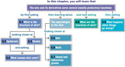
Overview
The integumentary system is the body's largest organ system, primarily serving as a protective barrier. It consists of the skin and its derivatives, including hair, nails, and glands. Understanding this system is essential for evaluating and treating skin injuries and diseases.
Structure of the Skin (Integument)
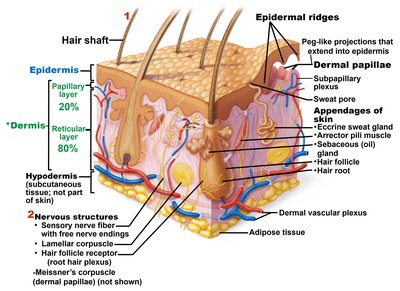
Main Layers of the Skin
Epidermis: The superficial, avascular layer composed of keratinized stratified squamous epithelium.
Dermis: The deeper, vascular layer made of dense irregular connective tissue, containing nerves, blood vessels, and appendages.
Hypodermis (Superficial Fascia): Subcutaneous layer deep to the skin, mainly adipose tissue; not part of the skin but anchors it to underlying structures and provides insulation.
Epidermis
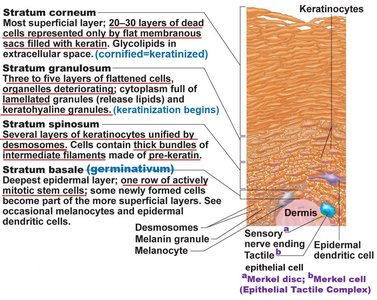
Layers of the Epidermis (from superficial to deep)
Stratum Corneum: 20–30 layers of dead, keratinized cells; provides a durable overcoat for protection.
Stratum Lucidum: Thin, clear layer found only in thick skin (palms, soles).
Stratum Granulosum: 1–5 layers where keratinization begins; cells accumulate keratohyaline and lamellar granules.
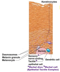
Stratum Spinosum: Several layers of keratinocytes unified by desmosomes; contains melanosomes and dendritic cells.
Stratum Basale: Deepest layer; single row of mitotically active stem cells; site of melanocytes and tactile cells.
Cell Types in the Epidermis
Keratinocytes: Produce keratin, the protein that gives skin its protective properties.
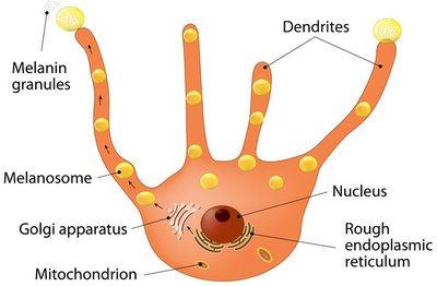
Melanocytes: Synthesize melanin pigment, which protects against UV radiation.
Dendritic (Langerhans) Cells: Immune cells that act as phagocytes and antigen-presenting cells.
Tactile Epithelial (Merkel) Cells: Associated with sensory nerve endings for touch sensation.
Dermis
Layers of the Dermis
Papillary Layer: Superficial areolar connective tissue with dermal papillae that project into the epidermis, containing capillary loops, Meissner's corpuscles (touch receptors), and free nerve endings.
Reticular Layer: Deeper, dense irregular connective tissue; provides strength, extensibility, and elasticity. Contains collagen and elastic fibers, hair follicles, and glands.
Dermal Papillae and Skin Markings
Dermal Papillae: Peg-like projections that increase surface area for exchange of gases, nutrients, and waste products between dermis and epidermis.
Friction Ridges: Formed by dermal and epidermal ridges; enhance gripping ability and create fingerprints.
Cleavage (Tension) Lines: Formed by parallel bundles of collagen fibers; important for surgical incisions.
Flexure Lines: Dermal folds at joints where skin cannot slide easily, causing deep creases.
Skin Color
Pigments Determining Skin Color
Melanin: Only pigment produced in the skin; ranges from reddish-yellow to brownish-black. All humans have similar numbers of melanocytes; color differences are due to type, amount, and retention of melanin.
Carotene: Yellow to orange pigment from plant products; accumulates in the stratum corneum and hypodermis.
Hemoglobin: Oxygenated hemoglobin in red blood cells gives a pinkish hue to fair skin.
Clinical Relevance of Skin Color
Cyanosis: Bluish tint due to poorly oxygenated hemoglobin.
Pallor: Pale color from emotional stress, anemia, or low blood pressure.
Erythema: Redness from fever, inflammation, or embarrassment.
Jaundice: Yellow cast from bilirubin accumulation (liver disorder).
Bruises: Clotted blood beneath the skin (hematomas).
Hair
Structure and Function
Composed of dead, keratinized cells (hard keratin).
Functions: Sensory reception, protection from trauma, heat loss, and sunlight.
Hair color is determined by the type and amount of melanin; red hair contains pheomelanin.
Hair types: Vellus (fine, pale) and terminal (coarse, long).
Hair Growth and Disorders
Growth cycles alternate between active and resting phases.
Alopecia: Hair thinning with age.
Male pattern baldness: Genetically determined, sex-linked.
Hirsutism: Excessive hair growth in females, often due to androgen excess.
Glands of the Skin
Sudoriferous (Sweat) Glands
Eccrine Glands: Most numerous; abundant on palms, soles, and forehead. Secrete hypotonic sweat for thermoregulation.
Apocrine Glands: Confined to axillary and anogenital areas; secrete sweat with fatty substances and proteins, which bacteria break down to produce body odor.
Modified Apocrine Glands: Ceruminous (earwax) and mammary (milk) glands.
Sebaceous (Oil) Glands
Widely distributed except in thick skin of palms and soles.
Secrete sebum (oily, acidic holocrine secretion) into hair follicles; lubricates and waterproofs skin, has bactericidal properties.
Functions of the Integumentary System
Major Functions
Protection: Physical, chemical, and biological barriers against pathogens, chemicals, and UV radiation.
Temperature Regulation: Sweat production and blood flow adjustments maintain body temperature.
Cutaneous Sensation: Sensory receptors detect pain, pressure, touch, and temperature.
Metabolic Functions: Synthesis of vitamin D precursor.
Blood Reservoir: Dermal blood vessels store about 5% of the body's blood volume.
Excretion: Elimination of nitrogenous wastes through sweat.
Skin Disorders and Clinical Considerations
Skin Cancer
Basal Cell Carcinoma: Most common, least malignant; arises from stratum basale.
Squamous Cell Carcinoma: Arises from keratinocytes of the stratum spinosum; can metastasize.
Melanoma: Cancer of melanocytes; highly metastatic and dangerous.
ABCDE Rule for Melanoma Detection: Asymmetry, Border irregularity, Color variation, Diameter >6mm, Evolving shape/size/color.
Burns
Classified by depth: First-degree (epidermis), Second-degree (epidermis and part of dermis), Third-degree (full thickness).
Severity estimated by the "Rule of Nines" (body divided into 11 areas, each 9% of total body area).
Critical burns: >25% body with second-degree, >10% with third-degree, or third-degree on hands, feet, face.
Immediate threats: Fluid loss, dehydration, electrolyte imbalance.
Additional info: The integumentary system is essential for homeostasis, immune defense, and sensory perception. Disorders can have systemic effects, highlighting the importance of skin health in overall physiology.
