 Back
BackThe Integumentary System: Structure, Function, and Clinical Relevance
Study Guide - Smart Notes
 Tailored notes based on your materials, expanded with key definitions, examples, and context.
The Integumentary System
Overview of the Integumentary System
The integumentary system is composed of the skin and its accessory structures, including hair, nails, and glands. It is the largest organ system in the human body, accounting for 10–15% of total body weight. The skin serves as a protective barrier and plays critical roles in sensation, thermoregulation, excretion, and synthesis of vitamin D.
Skin Structure
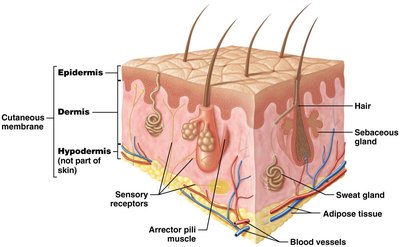
The skin consists of two main layers: the epidermis (keratinized stratified squamous epithelium) and the dermis (loose and dense irregular connective tissue). Deep to the dermis is the hypodermis (superficial fascia or subcutaneous fat), which is not part of the skin but anchors it to deeper structures and contains adipose tissue.
Epidermis: Avascular; receives nutrients via diffusion from the dermis.
Dermis: Vascular; provides blood supply, contains sensory receptors, and anchors the epidermis.
Hypodermis: Composed of loose connective tissue and adipose; highly vascular.
Accessory structures: Sweat glands, sebaceous glands, hair, nails.
Sensory receptors: Detect heat, cold, pain, and pressure.
Arrector pili muscles: Small bands of smooth muscle associated with hair follicles.
Functions of the Integumentary System
Major Functions
Protection: Shields against mechanical trauma, pathogens, and environmental hazards.
Sensation: Sensory receptors perceive changes in the internal and external environment.
Thermoregulation: Maintains stable internal temperature via negative feedback loops.
Excretion: Eliminates waste products and toxins through sweat.
Synthesis: Produces vitamin D (calcitriol), essential for calcium absorption.
Thermoregulation
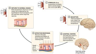
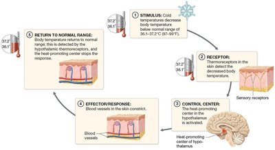
Thermoregulation is achieved through negative feedback mechanisms that adjust blood flow and sweat production in response to changes in body temperature.
When body temperature rises: Thermoreceptors detect the increase, the hypothalamus (control center) stimulates sweating and vasodilation of dermal vessels, leading to heat loss.
When body temperature falls: Thermoreceptors detect the decrease, the hypothalamus triggers vasoconstriction and shivering, reducing heat loss and generating heat.
Vitamin D Synthesis
UV light converts a precursor in the skin to vitamin D3 (cholecalciferol), which is then modified in the liver and kidneys to form calcitriol. Calcitriol is necessary for calcium absorption in the small intestine, which is essential for nerve function, muscle contraction, and bone health.
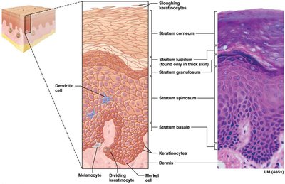
The Epidermis
Structure and Cell Types
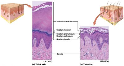
The epidermis is the most superficial region of the skin and is composed primarily of keratinocytes, which produce the protein keratin. The epidermis is organized into five distinct layers (strata):
Stratum basale (germinativum): Deepest layer; most metabolically active; site of keratinocyte mitosis and vitamin D precursor production.
Stratum spinosum: Still close to blood supply; metabolically active.
Stratum granulosum: Three to five layers of keratin-filled cells; provides water resistance.
Stratum lucidum: Narrow layer of clear, dead keratinocytes; found only in thick skin.
Stratum corneum: Outermost layer; several layers of dead, flattened cells; sloughed off mechanically.
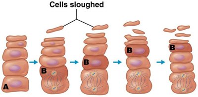
Keratinocyte Life Cycle
Keratinocytes are produced in the stratum basale and spinosum and migrate toward the surface, undergoing differentiation and eventually dying. The entire process takes 40–50 days.
Other Cells of the Epidermis
Dendritic (Langerhans) cells: Immune system phagocytes in the stratum spinosum.
Tactile (Merkel) cells: Sensory receptors for light touch, located in the stratum basale.
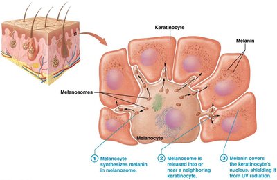
Melanocytes: Produce melanin pigment, located in the stratum basale.
Thick and Thin Skin
Thick skin: Contains all five epidermal layers, a thick stratum corneum, no hair follicles, and many sweat glands (e.g., palms, soles).
Thin skin: Lacks the stratum lucidum, has many hair follicles, sweat glands, and sebaceous glands.
Callus: Localized thickening of the stratum corneum due to repetitive pressure.
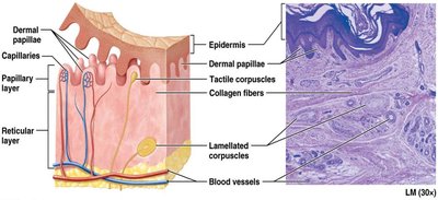
The Dermis
Structure and Layers
The dermis is a highly vascular layer deep to the epidermis, providing structural support and nourishment. It is composed of two layers:
Papillary layer: Loose connective tissue; contains dermal papillae, capillary loops, and tactile (Meissner) corpuscles for light touch.
Reticular layer: Dense irregular connective tissue; contains collagen and elastic fibers, lamellated (Pacinian) corpuscles for pressure and vibration, blood vessels, sweat glands, hair follicles, sebaceous glands, and adipose tissue.
Skin Markings and Wrinkles
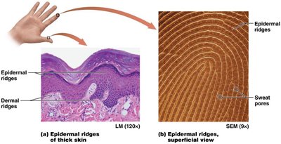
Epidermal ridges: Enhance gripping ability; form fingerprints; genetically determined patterns.
Wrinkles: Result from decreased collagen, elastic fibers, proteoglycans, and adipose tissue with age; accelerated by UV exposure and smoking.
Skin Color
Pigments and Clinical Relevance
Melanin: Produced by melanocytes; protects DNA from UV-induced mutations; amount produced determines skin tone.
Carotene: Yellow-orange pigment from diet; accumulates in the stratum corneum.
Hemoglobin: Red pigment in blood; skin coloration varies with blood flow.
Skin color can be a diagnostic tool:
Erythema: Increased blood flow (redness).
Pallor: Decreased blood flow (paleness).
Cyanosis: Low oxygenated blood (bluish color).
Jaundice: Yellow-orange color due to bilirubin buildup.

Melanin Synthesis and Function
Melanin is produced in melanosomes within melanocytes and transferred to keratinocytes, where it accumulates above the nucleus to shield DNA from UV radiation. Melanin synthesis increases with UV exposure but degrades after a few days, requiring continual replacement.
Common Pigmentation Variations
Freckle: Localized increase in melanin production.
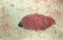
Mole (nevus): Proliferation of melanocytes.
Albinism: Genetic lack of tyrosinase enzyme, resulting in little or no melanin production.
Accessory Structures: Hair and Nails
Hair Structure and Function
Hair (pili) is derived from the epidermis and covers most of the body except thick skin, lips, and parts of the external genitalia. Hair protects against foreign substances, UV radiation, and mechanical trauma, and serves as a sensory structure.
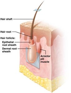
Shaft: Projects from the skin surface; composed of dead keratinized cells.
Root: Embedded in the dermis; surrounded by sensory neurons; contains the hair papilla (blood supply) and hair bulb (growth region).
Matrix: Actively dividing keratinocytes at the base of the root.
Hair follicle: Surrounds the root; composed of epithelial and dermal sheaths.
Arrector pili muscle: Smooth muscle causing "goosebumps" by making hair stand up.
Hair regions: Inner medulla (soft keratin), middle cortex (hard keratin), outer cuticle (overlapping keratinocytes).

Hair Pigment and Texture
Melanin: Determines hair color; blond hair has little melanin, black hair has much, red hair contains a special pigment with iron, gray/white hair results from decreased melanin with age.
Nails
Nails are composed of stratified squamous epithelium filled with hard keratin. They protect fingertips and aid in manipulation of objects.
Nail plate: Sits atop the nail bed.
Lunula: Crescent-shaped region at the base of the nail.
Eponychium: Cuticle.
Hyponychium: Stratum corneum under the free edge of the nail.
Glands of the Skin
Types of Glands
Sweat (sudoriferous) glands: Eccrine (widespread, watery secretion), apocrine (axillary/anal regions, functional at puberty, odoriferous, associated with hair follicle), ceruminous (ear canal, produce cerumen), mammary (produce milk).
Sebaceous glands: Secrete oily sebum; found in thin skin; provide a hydrophobic barrier.
Clinical Applications
Acne Vulgaris
Acne is caused by accumulation of sebum and dead cells in sebaceous glands, leading to comedones (blackheads) and, if infected, pustules (pimples). It is influenced by hormones and is common in adolescence.
Wounds and Burns
Wounds are disruptions in skin integrity and may involve the epidermis, dermis, or deeper tissues. Burns are classified by depth (first, second, third degree) and require different treatments.
Skin Cancer
Skin cancer is one of the most common cancers, often linked to UV exposure and carcinogens. It results from mutations that disrupt cell cycle control, leading to tumor formation and potential metastasis.
Type | Origin | Features |
|---|---|---|
Basal cell carcinoma | Stratum basale keratinocytes | Most common; rarely metastasizes |
Squamous cell carcinoma | Stratum spinosum keratinocytes | Second most common; may metastasize |
Malignant melanoma | Melanocytes | Most dangerous; high risk of metastasis |
The ABCDE rule helps distinguish malignant melanoma:
A: Asymmetry
B: Border irregularity
C: Color variation
D: Diameter > 6 mm
E: Evolving shape and size
*Additional info: This guide integrates foundational concepts and clinical relevance for the integumentary system, suitable for ANP college-level study and exam preparation.*
