 Back
BackThe Integumentary System: Structure, Function, and Clinical Relevance
Study Guide - Smart Notes
 Tailored notes based on your materials, expanded with key definitions, examples, and context.
The Integumentary System
Overview
The integumentary system is a complex organ system that serves as the body's primary barrier against the external environment. It consists of the skin, hair, nails, sweat glands, and sebaceous (oil) glands. This system plays crucial roles in protection, sensation, thermoregulation, and metabolic functions.
Skin: The largest organ, providing a protective covering.
Hair: Offers protection and sensory input.
Nails: Protects the distal phalanges.
Sweat glands: Regulate temperature and excrete wastes.
Sebaceous glands: Lubricate and protect skin and hair.
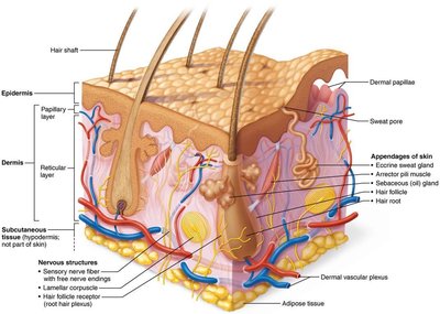
Skin Structure
Layers of the Skin
The skin is composed of two main layers and a subcutaneous region:
Epidermis: The superficial, avascular layer made of epithelial tissue.
Dermis: The deeper, vascular layer composed mainly of fibrous connective tissue.
Hypodermis (Subcutaneous layer): Not technically part of the skin, but anchors it to underlying structures and provides insulation and shock absorption via adipose tissue.
Epidermis
Cell Types in the Epidermis
The epidermis is primarily made of keratinized stratified squamous epithelium and contains four main cell types:
Keratinocytes: Produce keratin, the protein that gives skin its strength and waterproofing.
Melanocytes: Produce melanin pigment, protecting against UV radiation.
Dendritic (Langerhans) cells: Immune cells that patrol the epidermis.
Tactile (Merkel) cells: Sensory receptors for touch.
Layers of the Epidermis
The epidermis is organized into distinct layers (strata):
Stratum basale: Deepest layer, contains stem cells and melanocytes; site of active mitosis.
Stratum spinosum: Several layers thick, contains keratinocytes and dendritic cells; provides strength and flexibility.
Stratum granulosum: Cells flatten and keratinization begins; contains keratohyaline and lamellar granules.
Stratum lucidum: Present only in thick skin (palms, soles); consists of clear, dead keratinocytes.
Stratum corneum: Outermost layer; 20–30 rows of dead, keratinized cells providing protection.
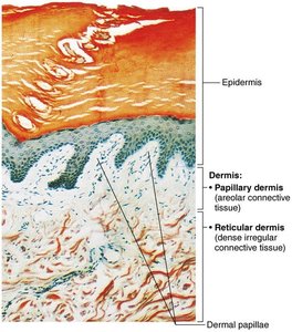
Dermis
Structure and Layers
The dermis is a strong, flexible connective tissue layer containing nerves, blood vessels, lymphatics, hair follicles, and glands. It is divided into two layers:
Papillary layer: Superficial, areolar connective tissue with dermal papillae that increase surface area for exchange and contain capillary loops and sensory receptors.
Reticular layer: Deep, dense irregular connective tissue with collagen and elastic fibers; provides strength, elasticity, and hydration.
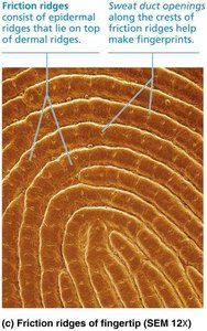
Friction Ridges
Dermal papillae in thick skin form friction ridges, which enhance gripping ability and create fingerprints.
Sweat duct openings along these ridges contribute to unique fingerprint patterns.
Skin Color
Pigments and Clinical Significance
Skin color is determined by three main pigments:
Melanin: Produced by melanocytes; shields DNA from UV damage.
Carotene: Yellow-orange pigment, accumulates in the stratum corneum and hypodermis.
Hemoglobin: Gives a pinkish hue to fair skin due to transparency.
Alterations in skin color can indicate disease states, such as cyanosis (blue), pallor (pale), erythema (red), jaundice (yellow), and hematomas (bruises).
Hair
Structure and Function
Hair consists of dead, keratinized cells and serves protective and sensory functions.
Shaft: Visible part above the skin; keratinization is complete.
Root: Embedded in the skin; keratinization ongoing.
Hair matrix: Actively dividing cells at the base of the follicle.
Arrector pili: Smooth muscle causing "goose bumps" by contracting and elevating the hair.
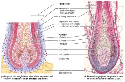
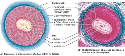
Parts of the Hair Shaft
Medulla: Central core with large cells and air spaces.
Cortex: Layers of flattened cells surrounding the medulla.
Cuticle: Outermost layer of overlapping single cells.
Hair color is determined by melanins produced in the follicle; red hair contains additional pheomelanin, while gray/white hair results from decreased melanin and air bubbles in the shaft.
Types and Growth of Hair
Vellus hair: Fine, pale body hair.
Terminal hair: Coarse, long hair found on scalp, eyebrows, and at puberty in axillary and pubic regions.
Hair growth is influenced by nutrition and hormones; follicles cycle between active and regressive phases.
Hair Thinning and Baldness
Alopecia: Hair thinning after age 40.
Male pattern baldness: Genetically determined, influenced by DHT (dihydrotestosterone).
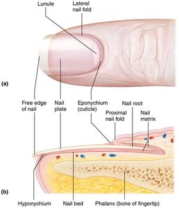
Nails
Structure and Clinical Relevance
Nails are scale-like modifications of the epidermis containing hard keratin. They protect the distal phalanges and consist of several parts:
Free edge, nail plate, root, nail bed, nail matrix (growth area), nail folds, eponychium (cuticle), hyponychium (under free edge).
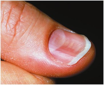
Nails appear pink due to underlying capillaries; the lunule is a white, thickened area of the matrix.
Abnormal nail color or shape can indicate disease (e.g., yellow nails for respiratory disorders, spoon nails for iron deficiency).
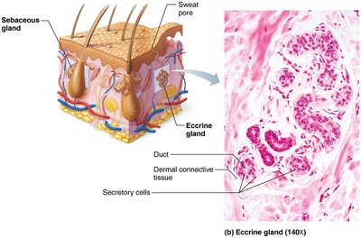
Cutaneous Glands
Sweat Glands
Sweat glands (sudoriferous glands) are distributed throughout the skin except for certain areas. There are two main types:
Eccrine (merocrine) glands: Most numerous, abundant on palms, soles, and forehead; function in thermoregulation and secrete watery sweat.
Apocrine glands: Confined to axillary and anogenital areas; secrete viscous sweat containing fatty substances and proteins, which bacteria break down to produce body odor.
Modified Apocrine Glands
Ceruminous glands: Secrete earwax (cerumen).
Mammary glands: Secrete milk.
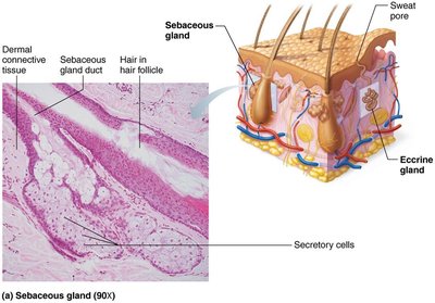
Sebaceous (Oil) Glands
Widely distributed except for thick skin; secrete sebum into hair follicles.
Activated at puberty by hormones; sebum lubricates and protects skin and hair, and has bactericidal properties.
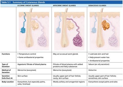
Summary Table: Cutaneous Glands
The following table summarizes the main types of cutaneous glands, their functions, methods of secretion, and locations:
Gland Type | Functions | Type of Secretion | Method of Secretion | Exits Duct At | Body Location |
|---|---|---|---|---|---|
Eccrine Sweat Glands | Temperature control; antibacterial properties | Hypotonic filtrate of blood plasma | Merocrine (exocytosis) | Skin surface | Everywhere, especially palms, soles, forehead |
Apocrine Sweat Glands | May act as sexual scent glands | Filtrate of blood plasma with added proteins and fatty substances | Merocrine (exocytosis) | Usually upper part of hair follicle; rarely skin surface | Mainly axillary and anogenital regions |
Sebaceous Glands | Lubricate skin and hair; antibacterial properties | Sebum (oil) | Holocrine | Usually upper part of hair follicle; sometimes skin surface | Everywhere except palms and soles |
Functions of Skin
Protection
The skin acts as a barrier through three mechanisms:
Chemical barrier: Secretes antimicrobial proteins, acid mantle, and melanin.
Physical barrier: Keratinized cells and glycolipids block water and most substances.
Biological barrier: Contains immune cells (dendritic cells, macrophages) and DNA absorbs UV radiation.
Body Temperature Regulation
Sweat glands produce insensible perspiration under normal conditions.
Increased sweat production (sensible perspiration) cools the body.
Dermal blood vessels constrict in cold environments to reduce heat loss.
Cutaneous Sensations
Sensory receptors in the skin detect touch, temperature, and pain.
Metabolic Functions
Skin synthesizes vitamin D, activates hormones, and produces collagenase.
Blood Reservoir
Skin can hold up to 5% of the body's blood volume and shunt blood as needed.
Excretion
Skin excretes nitrogenous wastes and regulates salt and water loss through sweating.
Clinical Relevance: Skin Disorders
Skin Cancer
Basal cell carcinoma: Least malignant, most common.
Squamous cell carcinoma: Can metastasize, good prognosis if treated.
Melanoma: Most dangerous, highly metastatic; early detection is critical (ABCD rule: Asymmetry, Border, Color, Diameter).
Burns
Burns are classified by severity: first-degree (epidermal), second-degree (epidermal and upper dermal), third-degree (entire skin thickness).
Serious burns are life-threatening due to dehydration and electrolyte imbalance.
The Rule of Nines is used to estimate fluid loss.
Developmental Aspects
Fetal to Adult Skin
By the 4th month, fetal skin is developed; lanugo coat and vernix caseosa protect the fetus.
Skin thickens and accumulates fat from infancy to adulthood; gland activity increases.
Aging leads to thinner, drier skin, decreased elasticity, increased risk of cancer, and hair thinning.
Preventive measures include UV protection, good nutrition, hydration, and hygiene.
References
Marieb, E. N., & Hoehn, K. (2023). Human anatomy and physiology (12th ed.). Pearson.
