 Back
BackThe Integumentary System: Structure, Function, and Clinical Aspects
Study Guide - Smart Notes
 Tailored notes based on your materials, expanded with key definitions, examples, and context.
The Integumentary System
Overview and Components
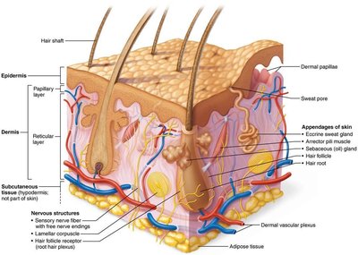
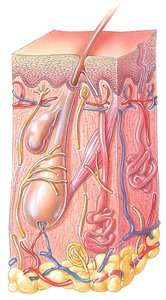
The integumentary system is the body's largest organ system, serving as a protective barrier and playing vital roles in sensation, thermoregulation, and metabolic processes. It consists of the skin, hair, nails, and various glands.
Skin: The primary organ, composed of multiple layers.
Hairs: Provide protection and sensory input.
Nails: Protect the distal phalanges and aid in manipulation.
Cutaneous (skin) glands: Include sweat (sudoriferous) and oil (sebaceous) glands.
Subcutaneous tissue (hypodermis): Anchors skin to underlying structures and provides insulation.
Structure of the Skin
Skin Layers
The skin is composed of two main layers and an underlying subcutaneous layer:
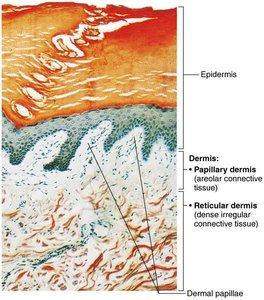
Epidermis: The outermost, avascular layer made of keratinized stratified squamous epithelium.
Dermis: The deeper, vascular layer composed of connective tissue, housing nerves, blood vessels, and glands.
Hypodermis (subcutaneous layer): Not technically part of the skin, but supports it with adipose and areolar tissue, providing insulation and shock absorption.
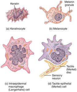
Epidermal Cell Types
The epidermis contains four main cell types, each with specialized functions:
Keratinocytes: Produce keratin, a protein that strengthens and waterproofs the skin.
Melanocytes: Synthesize melanin pigment, protecting against UV radiation.
Dendritic (Langerhans) cells: Immune cells that detect and process antigens.
Tactile epithelial (Merkel) cells: Sensory receptors for touch.
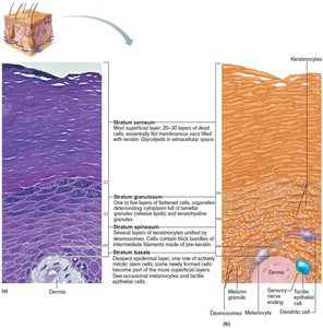
Layers of the Epidermis
The epidermis is organized into distinct layers (strata), with thick skin (palms, soles) containing five layers and thin skin containing four:
Stratum basale: Deepest, mitotically active layer; contains stem cells and melanocytes.
Stratum spinosum: Several layers of keratinocytes connected by desmosomes; contains dendritic cells and melanosomes.
Stratum granulosum: Cells flatten, organelles disintegrate, and keratinization begins; contains keratohyaline and lamellar granules.
Stratum lucidum: Present only in thick skin; thin, clear layer of dead keratinocytes.
Stratum corneum: Outermost layer; 20–30 layers of dead, keratinized cells providing a durable overcoat.
Dermis
The dermis is a strong, flexible connective tissue layer divided into two regions:
Papillary dermis: Superficial areolar connective tissue with dermal papillae containing capillaries, nerve endings, and touch receptors.
Reticular dermis: Deeper, dense irregular connective tissue with collagen and elastic fibers, providing strength and elasticity.
Dermal Modifications
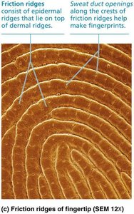
Friction ridges: Form fingerprints; enhance grip and tactile sensitivity.
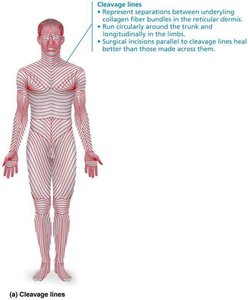
Cleavage (tension) lines: Indicate orientation of collagen fibers; important for surgical incisions.
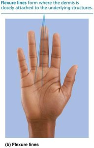
Flexure lines: Dermal folds at joints, allowing skin to accommodate movement.
Clinical Aspects
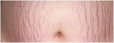
Striae (stretch marks): Result from dermal tearing due to rapid stretching.
Blisters: Fluid-filled pockets separating epidermal and dermal layers, often due to acute trauma.
Basis of Skin Color
Pigments
Melanin: Produced by melanocytes; protects DNA from UV damage. Varies in amount and type, causing differences in skin color.
Carotene: Yellow to orange pigment, most visible in palms and soles; can be converted to vitamin A.
Hemoglobin: Oxygenated pigment in red blood cells; gives fair skin a pinkish hue.
Alterations in skin color can indicate disease (e.g., cyanosis, jaundice, pallor, erythema, bruising).
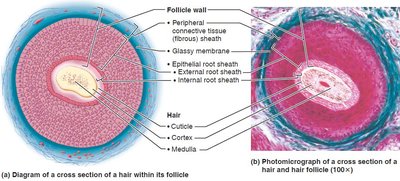
Hair
Structure and Function
Hair consists of dead, keratinized cells and serves protective and sensory functions. It is absent from palms, soles, lips, nipples, and parts of external genitalia.
Shaft: Visible part above the skin; keratinization is complete.
Root: Embedded in the skin; keratinization ongoing.
Hair follicle: Extends from epidermis into dermis; contains the hair bulb, matrix, and associated structures.

Types of Hair
Lanugo: Fine, unpigmented hair on fetus.
Vellus hair: Fine, pale body hair of children and adult females.
Terminal hair: Coarse, pigmented hair on scalp, eyebrows, and after puberty in axillary and pubic regions.
Hair color is determined by the type and amount of melanin produced by follicular melanocytes.
Hair Growth and Disorders
Hair growth is influenced by nutrition and hormones, cycling through growth, regression, and resting phases.
Alopecia: Hair thinning with age.
Male pattern baldness: Genetically determined, androgen-influenced hair loss.
Hirsutism: Excessive hair growth in females, often due to androgen excess.
Telogen effluvium: Sudden hair thinning due to stress or physiological changes.
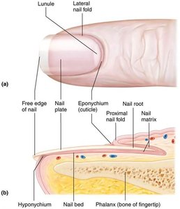
Nails
Structure and Function
Nails are scale-like modifications of the epidermis, composed of hard keratin. They protect the distal phalanges and aid in manipulation.
Nail plate: Visible attached part of the nail.
Free edge: Distal portion that extends beyond the finger or toe.
Nail root: Proximal part embedded under the skin.
Nail matrix: Site of nail growth.
Eponychium (cuticle): Skin fold overlapping the nail.
Hyponychium: Area under the free edge.
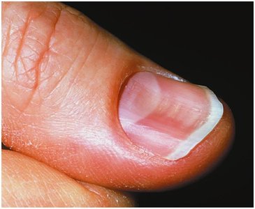
Abnormal nail appearance can indicate systemic diseases (e.g., spoon nails in iron deficiency, Beau's lines in severe illness).
Cutaneous (Skin) Glands
Types of Glands
Sudoriferous (sweat) glands: Eccrine and apocrine types; involved in thermoregulation and scent production.
Sebaceous (oil) glands: Secrete sebum to lubricate and waterproof skin and hair.
Ceruminous glands: Produce earwax.
Mammary glands: Produce milk.
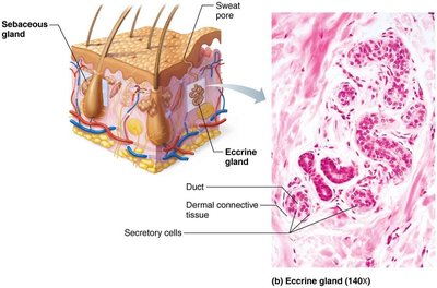
Eccrine (Merocrine) Sweat Glands
Most numerous; abundant on palms, soles, and forehead.
Secrete watery sweat for thermoregulation.
Regulated by the sympathetic nervous system.
Apocrine Sweat Glands
Confined to axillary and anogenital areas.
Secrete viscous, protein-rich sweat; responsible for body odor when decomposed by bacteria.
Include modified types: ceruminous (earwax) and mammary (milk) glands.

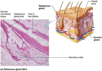
Sebaceous (Oil) Glands
Widely distributed except on palms and soles.
Secrete sebum via holocrine secretion; lubricates and protects skin and hair.
Activity increases at puberty due to androgens.
Clinical Aspects of Glands
Acne: Inflammation of sebaceous glands, often due to bacterial infection.
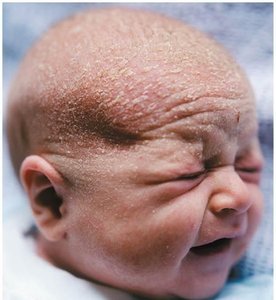
Seborrhea (cradle cap): Overactive sebaceous glands in infants, causing flaky scalp lesions.
Functions of the Skin
Major Functions
Protection: Physical, chemical, and biological barriers against pathogens, chemicals, and UV radiation.
Body temperature regulation: Sweat production and blood flow adjustments maintain thermal homeostasis.
Cutaneous sensations: Sensory receptors detect touch, temperature, pain, and pressure.
Metabolic functions: Synthesis of vitamin D, activation of hormones, and breakdown of carcinogens.
Blood reservoir: Stores up to 5% of the body's blood volume.
Excretion: Eliminates nitrogenous wastes and excess salts via sweat.
Skin Cancer and Burns
Skin Cancer
Skin cancer is primarily caused by UV exposure and chronic irritation. There are three major types:
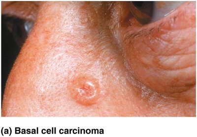
Basal cell carcinoma: Most common, least malignant; arises from stratum basale.
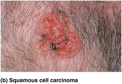
Squamous cell carcinoma: Arises from keratinocytes of stratum spinosum; can metastasize.
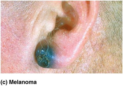
Melanoma: Cancer of melanocytes; highly metastatic and dangerous.
The ABCD rule helps identify melanoma: Asymmetry, Border irregularity, Color variation, Diameter >6 mm.
Burns
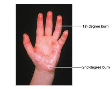
Burns are classified by depth and severity:
First-degree: Epidermal damage only; redness and pain.
Second-degree: Epidermal and upper dermal damage; blisters form.
Third-degree: Full-thickness damage; skin may appear white, red, or blackened; nerve endings destroyed.
Burns are critical if >25% of body has second-degree burns, >10% has third-degree burns, or if face, hands, or feet are involved. Treatment includes fluid replacement, infection control, and skin grafting.
Developmental Aspects of the Integumentary System
Fetal Development
By the 4th month, fetal skin is developed.
Lanugo coat (fine hair) appears in the 5th–6th month.
Vernix caseosa (sebaceous secretion) protects fetal skin in amniotic fluid.
Changes from Infancy to Adulthood
Skin thickens, subcutaneous fat increases, and gland activity rises during growth.
Optimal appearance in 20s–30s; aging leads to thinning, dryness, decreased elasticity, and increased cancer risk.
Prevention of aging: UV protection, good nutrition, hydration, and hygiene.
