 Back
BackThe Integumentary System: Structure, Function, and Clinical Aspects
Study Guide - Smart Notes
 Tailored notes based on your materials, expanded with key definitions, examples, and context.
The Integumentary System
Overview
The integumentary system is the body's largest organ system, serving as a protective barrier and playing vital roles in sensation, thermoregulation, and metabolic processes. It consists of the skin, sweat glands, oil glands, hair, nails, and subcutaneous tissue.
Skin: The primary organ, composed of two main layers (epidermis and dermis) and an underlying subcutaneous layer.
Appendages: Includes hair, nails, and cutaneous glands (sweat and sebaceous glands).
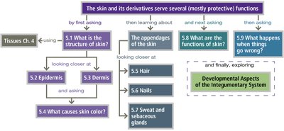
5.1 Skin Structure
Layers of the Skin
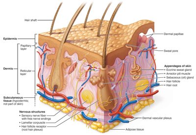
The skin is composed of two main layers and an associated subcutaneous layer:
Epidermis: Outermost, avascular, composed of keratinized stratified squamous epithelium.
Dermis: Deeper, vascular, made of dense connective tissue.
Subcutaneous Tissue (Hypodermis): Not part of the skin proper, but anchors skin to underlying structures, provides insulation and shock absorption.
5.2 The Epidermis
Cell Types in the Epidermis
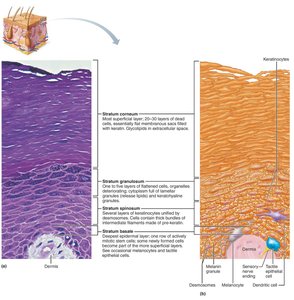
Keratinocytes: Produce keratin, the protein that gives skin its protective properties; most abundant cell type.
Melanocytes: Produce melanin pigment, which protects against UV radiation.
Dendritic (Langerhans) Cells: Immune cells that patrol the epidermis and activate immune responses.
Tactile (Merkel) Cells: Sensory receptors for touch, located at the epidermal-dermal junction.
Layers (Strata) of the Epidermis
The epidermis is organized into distinct layers:
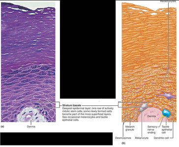
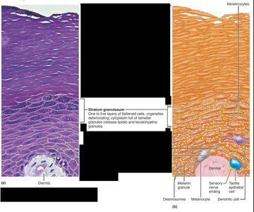
Stratum Basale: Deepest, single row of mitotically active stem cells; contains melanocytes.
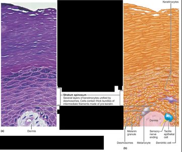
Stratum Spinosum: Several layers thick, contains pre-keratin filaments and abundant dendritic cells.
Stratum Granulosum: Cells flatten, organelles disintegrate, keratinization begins, and glycolipids are released for waterproofing.
Stratum Lucidum: Thin, clear layer found only in thick skin (palms, soles).
Stratum Corneum: Outermost, 20–30 layers of dead, keratinized cells providing protection.
5.3 The Dermis
Structure and Function
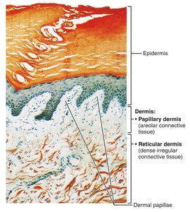
The dermis is a strong, flexible connective tissue layer housing blood vessels, nerves, hair follicles, and glands. It is divided into two layers:
Papillary Dermis: Superficial, areolar connective tissue with dermal papillae that contain capillaries, nerve endings, and touch receptors.
Reticular Dermis: Deep, dense irregular connective tissue with collagen and elastic fibers, providing strength and elasticity.
Dermal Modifications
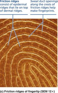
Friction Ridges: Form fingerprints, enhance grip and tactile sensation.
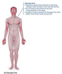
Cleavage (Tension) Lines: Orientation of collagen fibers; important for surgical incisions.
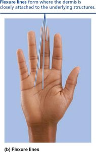
Flexure Lines: Dermal folds at joints, visible on palms and fingers.
5.4 Skin Color
Pigments Determining Skin Color
Melanin: Only pigment produced in the skin; protects DNA from UV damage. Varies in amount and type (reddish-yellow to brown-black).
Carotene: Yellow to orange pigment, most visible in palms and soles; can be converted to vitamin A.
Hemoglobin: Pinkish hue in fair skin due to low melanin and visible blood flow.
Alterations in skin color can indicate disease (e.g., cyanosis, jaundice, pallor, erythema, bruising).
5.5 Hair
Structure and Function
Hair (Pili): Dead, keratinized cells produced by hair follicles; functions in protection, sensation, and filtration.
Regions: Root (in scalp, keratinization ongoing) and shaft (above scalp, keratinization complete).
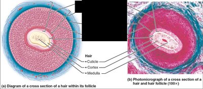
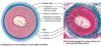
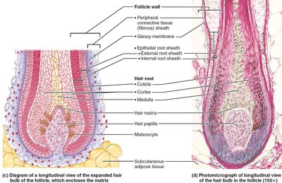
Hair Shaft Layers: Medulla (core), cortex (middle), cuticle (outermost).
Hair Follicle Structure
Follicle Wall: Peripheral connective tissue sheath, glassy membrane, epithelial root sheath.
Hair Bulb: Expanded base with hair matrix (growth zone) and papilla (nutrient supply).
Arrector Pili: Smooth muscle causing "goose bumps."
Types and Growth of Hair
Vellus Hair: Fine, pale body hair of children and adult females.
Terminal Hair: Coarse, long hair (scalp, eyebrows, axillary, pubic regions, face/neck in males).
Growth cycles alternate between active and resting phases; affected by nutrition and hormones.
5.6 Nails
Structure and Function
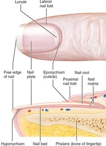
Nails: Scale-like modifications of the epidermis containing hard keratin; protect distal phalanges.
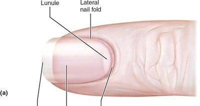
Parts: Root, nail plate (body), free edge, nail bed, nail matrix (growth zone), nail folds, cuticle (eponychium), hyponychium.
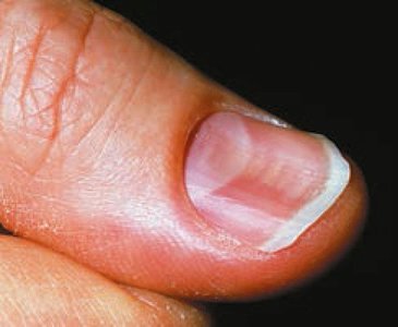
Abnormal nail appearance can indicate systemic disease (e.g., spoon nails in iron deficiency, Beau's lines in severe illness).
5.7 Sweat and Sebaceous Glands
Sweat (Sudoriferous) Glands
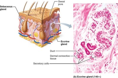
Eccrine (Merocrine) Glands: Most numerous, abundant on palms, soles, forehead; function in thermoregulation; secrete hypotonic sweat (99% water, salts, wastes).
Apocrine Glands: Axillary and anogenital areas; secrete viscous, protein-rich sweat into hair follicles; function begins at puberty; may act as scent glands.
Modified Apocrine Glands: Ceruminous (earwax) and mammary (milk) glands.
Sebaceous (Oil) Glands
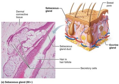
Widely distributed except on palms and soles; secrete sebum (oily, bactericidal, softens hair and skin).
Most develop from hair follicles; holocrine secretion method.
Summary Table: Cutaneous Glands
Eccrine Sweat Glands | Apocrine Sweat Glands | Sebaceous Glands | |
|---|---|---|---|
Function | Temperature control, some antibacterial properties | May act as sexual scent glands | Lubricate skin and hair, prevent water loss, antibacterial |
Type of Secretion | Hypotonic filtrate of blood | Filtrate of blood with proteins and fatty substances | Sebum (oily secretion) |
Method of Secretion | Merocrine (exocytosis) | Merocrine (exocytosis) | Holocrine |
Secretion Exit | Skin surface | Upper part of hair follicle | Upper part of hair follicle or skin surface |
Location | Everywhere, especially palms, soles, forehead | Axillary and anogenital regions | Everywhere except palms and soles |
5.8 Functions of Skin
Main Functions
Protection: Chemical (acid mantle, melanin), physical (keratinized cells), and biological (immune cells) barriers.
Body Temperature Regulation: Sweat production and blood vessel dilation/constriction.
Cutaneous Sensations: Touch, pressure, pain, and temperature receptors.
Metabolic Functions: Vitamin D synthesis, activation of hormones, collagenase production.
Blood Reservoir: Holds up to 5% of blood volume.
Excretion: Removal of nitrogenous wastes, salt, and water via sweat.
5.9 Skin Cancer and Burns
Skin Cancer
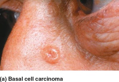
Basal Cell Carcinoma: Most common, least malignant; arises from stratum basale; high cure rate with excision.
Squamous Cell Carcinoma: Arises from keratinocytes of stratum spinosum; can metastasize; good prognosis if treated early.
Melanoma: Cancer of melanocytes; most dangerous due to high metastatic potential and resistance to therapy.
Burns
First-degree: Epidermal damage only; redness, pain, swelling.
Second-degree: Epidermal and upper dermal damage; blisters form.
Third-degree: Full-thickness burn; skin appears gray-white, cherry red, or blackened; not painful (nerve endings destroyed); requires grafting.
Severity is estimated using the "rule of nines" to calculate fluid loss and extent of burns.
Developmental Aspects of the Integumentary System
Embryonic to Adult Development
Skin develops from ectoderm (epidermis) and mesoderm (dermis, subcutaneous tissue).
At birth, skin is covered with vernix caseosa (protective substance) and lanugo (fine hair).
With age, skin thickens, glands become more active, and hair patterns change.
Aging leads to thinning, dryness, decreased elasticity, and increased risk of cancer.
