 Back
The Integumentary System: Structure, Function, and Clinical Aspects
Study Guide - Smart Notes
 Tailored notes based on your materials, expanded with key definitions, examples, and context.
The Integumentary System
Overview and Functions
The integumentary system consists of the skin and its accessory structures, including hair, nails, and glands. It is the largest organ system in the human body, accounting for approximately 16% of total body weight. The integumentary system serves as the body's first line of defense and is essential for maintaining homeostasis.
Protection: Acts as a barrier against mechanical injury, pathogens, and harmful chemicals.
Prevention of Water Loss: Minimizes dehydration by reducing water evaporation.
Sensory Input: Contains receptors for touch, pressure, pain, and temperature.
Thermoregulation: Regulates body temperature through sweat production and blood flow.
Vitamin D Synthesis: Initiates the synthesis of vitamin D upon exposure to UV radiation.
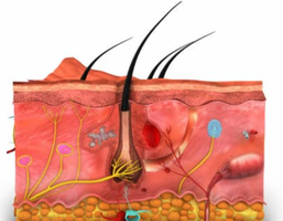
Layers of the Skin
Epidermis
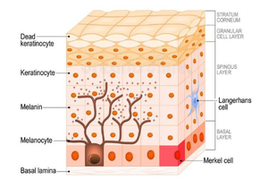
The epidermis is the outermost layer of the skin, composed of keratinized stratified squamous epithelium. It provides a tough, protective barrier and is avascular, relying on the underlying dermis for nutrient supply. The epidermis is organized into several layers, each with specialized functions.
Barrier Function: Protects against environmental hazards such as microbes, chemicals, and dehydration.
Cell Renewal: New cells form at the base and migrate upward, replacing older cells.
Dermis
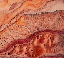
The dermis lies beneath the epidermis and is composed of dense connective tissue. It provides structural strength, flexibility, and houses blood vessels, nerves, hair follicles, and glands. The dermis is divided into two layers:
Papillary Layer: Thin, made of loose connective tissue; contains dermal papillae that increase surface area for nutrient exchange and strengthen attachment to the epidermis.
Reticular Layer: Thicker, made of dense connective tissue; contains collagen and elastin fibers, hair follicles, glands, and deep pressure receptors.
Hypodermis (Subcutaneous Layer)
The hypodermis is the deepest layer, consisting of loose connective tissue and adipose tissue. It cushions internal organs, insulates the body, and serves as an energy reserve. The hypodermis is also a common site for subcutaneous injections.
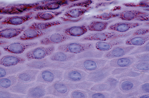
Cell Types and Layers of the Epidermis
Major Cell Types
Keratinocytes: Most abundant; produce keratin for strength and waterproofing.
Melanocytes: Produce melanin, which protects against UV radiation.
Merkel Cells: Sensory receptors for touch.
Basal Cells: Stem cells in the stratum basale; give rise to new keratinocytes.
Langerhans Cells: Immune cells that detect pathogens and participate in skin defense.
Layers of the Epidermis (Deep to Superficial)
Stratum Basale: Single layer of basal cells and melanocytes; site of cell division.
Stratum Spinosum: Several layers of keratinocytes connected by desmosomes.
Stratum Granulosum: Keratinocytes flatten, fill with keratin, and begin to die.
Stratum Lucidum: Present only in thick skin (palms, soles); clear layer of dead keratinocytes.
Stratum Corneum: Outermost layer of dead, flattened keratinocytes; forms a protective barrier.
Accessory Structures of the Skin
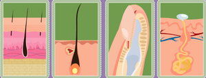
Hair
Hair is produced in follicles and consists of the bulb, root, and shaft. The hair matrix contains actively dividing cells, while the hair papilla supplies blood. Hair growth occurs in cycles: anagen (growth), catagen (regression), and telogen (resting). Arrector pili muscles cause hair to stand up (goosebumps).
Nails
Nail Matrix: Site of cell division at the base of the nail.
Nail Body: Visible portion of the nail.
Lunula: Pale crescent at the base.
Cuticle/Eponychium: Protective layer at the base.
Glands
Sebaceous Glands: Produce sebum for lubrication; associated with hair follicles.
Sweat Glands (Sudoriferous):
Eccrine: Widespread, produce watery sweat for cooling.
Apocrine: Found in armpits/groin; produce thicker sweat, associated with hair follicles.
Skin Pigmentation
Skin color is determined by the amount and type of melanin (produced by melanocytes), carotene (from diet), and hemoglobin (in blood). Melanin is packaged into melanosomes and transferred to keratinocytes, providing UV protection. Variations in these pigments account for differences in skin color among individuals.
Functions: Thermoregulation and Vitamin D Synthesis
Thermoregulation
Blood vessels in the dermis constrict or dilate to conserve or release heat.
Eccrine sweat glands secrete sweat for evaporative cooling.
Arrector pili muscles raise hair to trap heat.
Vitamin D Synthesis
UV radiation triggers the production of vitamin D in the skin.
Vitamin D is essential for calcium absorption and bone health.
Deficiency can lead to rickets, characterized by soft, weak bones and skeletal abnormalities.
Diseases, Disorders, and Injuries
Skin Cancer
Basal Cell Carcinoma: Most common; originates in the stratum basale; rarely metastasizes.
Squamous Cell Carcinoma: Originates in the stratum spinosum; can metastasize if untreated.
Melanoma: Originates from melanocytes; highly aggressive and prone to metastasis.
Burns
First Degree: Affects only the epidermis; redness and pain.
Second Degree: Involves epidermis and dermis; blisters form.
Third Degree: Full-thickness; may require grafting.
Fourth Degree: Extends into muscle or bone.
Skin Surface Disorders and Conditions
Stretch Marks: Tears in dermal collagen from rapid growth or stretching.
Callus: Thickened epidermis due to repeated friction.
Corn: Localized thickened skin over bony prominences.
Eczema: Chronic inflammation; red, itchy skin.
Acne: Blocked sebaceous glands, often with infection.
Vitiligo: Loss of melanocytes, causing depigmented patches.
Albinism: Genetic absence of melanin; increases UV sensitivity.
Rickets: Soft, weak bones due to vitamin D deficiency.
Summary Table: Layers and Functions of the Skin
Layer | Main Components | Functions |
|---|---|---|
Epidermis | Keratinocytes, melanocytes, Langerhans cells, Merkel cells | Protection, barrier, sensation, pigmentation |
Dermis | Collagen, elastin, blood vessels, nerves, glands, hair follicles | Strength, flexibility, nutrient supply, sensation, thermoregulation |
Hypodermis | Adipose tissue, loose connective tissue | Cushioning, insulation, energy storage |
Key Terms and Definitions
Keratin: A tough, fibrous protein produced by keratinocytes.
Melanin: Pigment responsible for skin color and UV protection.
Desmosomes: Cell junctions that connect keratinocytes in the epidermis.
Fibroblasts: Cells in the dermis that produce collagen and elastin fibers.
Sebum: Oily substance produced by sebaceous glands for lubrication.
Additional info:
Meissner corpuscles (in dermal papillae) detect light touch; Pacinian corpuscles (in reticular layer) detect pressure and vibration.
Skin renewal cycle is approximately 4 weeks.
