 Back
The Integumentary System: Structure, Function, and Clinical Aspects
Study Guide - Smart Notes
 Tailored notes based on your materials, expanded with key definitions, examples, and context.
The Integumentary System
Overview and Components
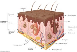
The integumentary system is the largest organ system of the body, primarily composed of the skin (cutaneous membrane), along with its derivatives such as hair, nails, sebaceous (oil) glands, and sudoriferous (sweat) glands. This system serves as the body's first line of defense and plays a critical role in homeostasis.
Skin: Largest organ, forms a protective barrier.
Nails: Scalelike modifications of the epidermis, composed of hard keratin.
Hair: Columns of dead keratinized cells, important for protection and sensation.
Glands: Sebaceous (oil) and sudoriferous (sweat) glands regulate moisture and temperature.
Functions of the Integument
Protection: Shields underlying tissues from mechanical damage, pathogens, and UV radiation.
Prevents Water Loss: Keratin and glycolipids reduce water evaporation.
Temperature Regulation: Sweat glands and blood vessels help maintain body temperature.
Metabolic Regulation: Synthesis of vitamin D upon exposure to sunlight.
Immune Defense: Langerhans cells detect and fight pathogens.
Sensory Reception: Nerve endings detect touch, pain, and temperature.
Excretion: Sweat glands excrete waste products.
Structure of the Skin
Major Regions
Epidermis: Superficial, avascular, composed of keratinized stratified squamous epithelium.
Dermis: Deep to the epidermis, contains connective tissue, blood vessels, nerves, and appendages.
Epidermis: Layers and Cells
The epidermis consists of five distinct layers (strata) in thick skin and four in thin skin. Each layer has specialized cells and functions.
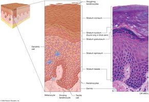
Keratinocytes: Produce keratin, the main structural protein.
Melanocytes: Produce melanin, which protects against UV radiation.
Langerhans (Dendritic) Cells: Immune cells that detect pathogens.
Merkel (Tactile) Cells: Sensory receptors for touch.
Layers of the Epidermis (from deep to superficial)
Stratum Basale: Single layer of cuboidal/columnar cells; site of mitosis; contains melanocytes and Merkel cells.
Stratum Spinosum: Several layers of keratinocytes; contains Langerhans cells; cells connected by desmosomes.
Stratum Granulosum: 2-5 layers of flattened cells; keratinization begins; contains keratohyaline and lamellar granules.
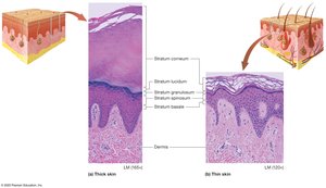
Stratum Lucidum: Thin, translucent layer found only in thick skin (palms, soles); consists of dead keratinocytes.
Stratum Corneum: 20-30 layers of dead, keratinized cells; provides barrier and protection.
Thick vs. Thin Skin
Thick Skin: Found on palms and soles; has all five strata; lacks hair follicles and sebaceous glands.
Thin Skin: Covers most of the body; lacks stratum lucidum; contains hair follicles and sebaceous glands.
Mnemonic for Epidermal Layers
Brilliant Basale
Studying Spinosum
Gives Granulosum
Loads of Lucidum
Confidence Corneum
Dermis
The dermis is a connective tissue layer beneath the epidermis, providing strength, flexibility, and nourishment to the skin. It contains two layers:
Papillary Layer: Superficial, composed of areolar connective tissue; contains dermal papillae, capillaries, and sensory receptors.
Reticular Layer: Deep, dense irregular connective tissue; contains collagen and elastic fibers, blood vessels, glands, and hair follicles.
Special Features
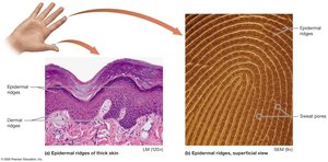
Dermal Papillae: Projections that increase surface area and form fingerprints.
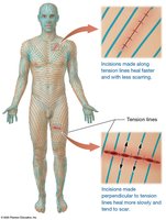
Cleavage (Tension) Lines: Orientation of collagen fibers; important for surgical incisions.
Flexure Lines: Dermal folds at joints (e.g., palms).
Stretch Marks: Tears in the dermis due to rapid stretching.
Hypodermis (Subcutaneous Layer)
The hypodermis is not part of the skin but lies deep to the dermis. It anchors the skin to underlying tissues and stores fat.
Composed of adipose and areolar connective tissue.
Functions in insulation, energy storage, and shock absorption.
Accessory Structures
Hair
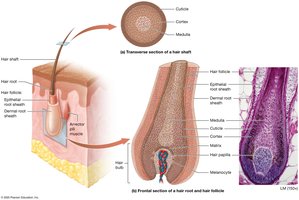
Hair is composed of dead, keratinized epithelial cells and serves protective and sensory functions.
Hair Shaft: Visible part above the skin surface.
Hair Root: Embedded in the skin, surrounded by the follicle.
Hair Bulb: Expanded base of the root; site of hair growth.
Dermal Papilla: Contains blood vessels and nerves for nourishment.
Piloerector (Arrector Pili) Muscle: Smooth muscle that causes hair to stand (goosebumps).
Types of Hair
Vellus Hair: Fine, pale hair on children and adult females.
Terminal Hair: Coarse, pigmented hair (scalp, eyebrows, etc.).
Nails
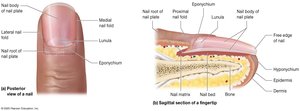
Nails are protective coverings on the dorsal surface of fingers and toes, composed of hard keratin.
Free Edge: Distal, visible part.
Body: Main visible portion.
Root: Proximal part embedded in skin.
Nail Folds: Skin folds overlapping the nail.
Eponychium: Cuticle, protects the nail matrix.
Glands of the Integumentary System
Classification of Glands
Endocrine Glands: Secrete hormones into the bloodstream.
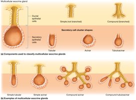
Exocrine Glands: Secrete products onto body surfaces or into ducts.
Types of Exocrine Glands in Skin
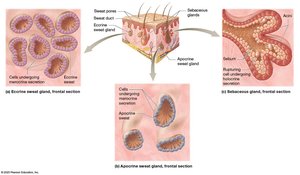
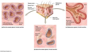
Sudoriferous (Sweat) Glands: Merocrine (eccrine) and apocrine types.
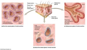
Sebaceous (Oil) Glands: Secrete sebum into hair follicles.
Ceruminous Glands: Produce earwax in the external ear canal.
Lactiferous Glands: Modified sweat glands producing milk.
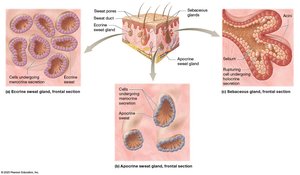
Merocrine (Eccrine) Sweat Glands
Most numerous; found on palms, soles, and forehead.
Secrete watery sweat for thermoregulation.
Apocrine Sweat Glands
Located in axillary and genital areas; become active at puberty.
Secrete a thicker fluid into hair follicles; responsible for body odor.
Sebaceous (Oil) Glands
Associated with hair follicles; secrete sebum to lubricate skin and hair.
Absent on palms and soles.
Skin Color and Clinical Aspects
Determinants of Skin Color
Melanin: Pigment produced by melanocytes; protects against UV radiation.
Hemoglobin: Oxygen-carrying pigment in blood; gives skin a pinkish hue.
Carotene: Yellow-orange pigment from diet; accumulates in stratum corneum.
Albinism: Genetic lack of melanin production.
Vitiligo: Loss of melanocytes in patches.
Nevus (Mole): Benign overgrowth of melanocytes.
Diagnostic Uses of the Integument
Cyanosis: Blue skin due to low oxygen.
Jaundice: Yellow skin from excess bilirubin.
Pallor: Pale skin from reduced blood flow.
Erythema: Red skin from increased blood flow.
Rashes/Hives: Indicate allergic reactions.
Wounds, Burns, and Skin Cancer
Wounds
Disruption of skin integrity; may involve epidermis, dermis, or deeper tissues.
Lacerations: Cuts or tears, often repaired with sutures.
Burns
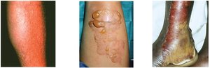
First-Degree: Affects only epidermis; redness and pain.
Second-Degree: Involves epidermis and part of dermis; blisters.
Third-Degree: Destroys epidermis and dermis; may extend to deeper tissues.
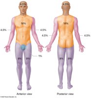
Rule of Nines
Used to estimate the percentage of body surface area affected by burns for fluid resuscitation.
Skin Cancer
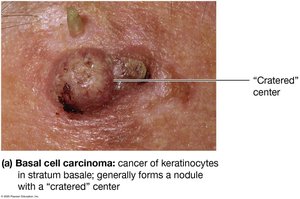
Basal Cell Carcinoma: Most common; arises from stratum basale; rarely metastasizes.
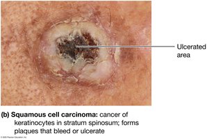
Squamous Cell Carcinoma: Arises from stratum spinosum; may metastasize.
Malignant Melanoma: Cancer of melanocytes; highly metastatic; prognosis depends on early detection.
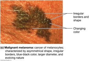
ABCDE Rule for Melanoma
Asymmetry: Two sides do not match.
Border irregularity: Edges are notched or blurred.
Color: Multiple or changing colors.
Diameter: Greater than 6 mm.
Evolving: Changes in size, shape, or color.
Tissue Repair and Aging
Tissue Repair
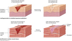
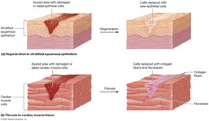
Regeneration: Replacement of damaged cells with the same type; restores function.
Fibrosis: Replacement with scar tissue (collagen); function may be lost.
Aging and the Integumentary System
Wrinkling and decreased elasticity.
Reduced immune responsiveness.
Dehydration and cracking of skin.
Decreased sweat and oil production.
Loss of subcutaneous fat and thinning of skin.
Slower growth of hair and nails; increased brittleness.
References: Amerman, E. C., 2019; Marieb, E. N., 2010; McKinley, M. and O’Loughlin, V. D., 2008; Seeley, R. R., et al., 2010; Tortora and Derrickson, 2014; Wise, E., 2008.
