 Back
BackThe Integumentary System: Structure, Function, and Clinical Relevance
Study Guide - Smart Notes
 Tailored notes based on your materials, expanded with key definitions, examples, and context.
The Integumentary System
Overview and Major Components
The integumentary system is the largest organ system of the human body, comprising approximately 16% of total body weight and covering 1.5 to 2 m2 of surface area. It consists of two primary parts: the cutaneous membrane (skin) and accessory structures. The cutaneous membrane is further divided into the outer epidermis and the inner dermis, while accessory structures include hair, exocrine glands, and nails.
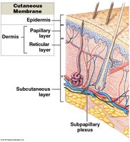
Cutaneous membrane: Composed of the epidermis (superficial epithelium) and dermis (connective tissue).
Accessory structures: Originate in the dermis and extend through the epidermis to the surface (hair, glands, nails).
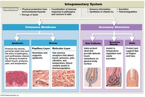
Functions of the Integumentary System
The integumentary system performs several vital functions essential for homeostasis and protection:
Protection: Shields underlying tissues and organs from mechanical damage, pathogens, and chemical exposure.
Excretion: Removes salts, water, and organic wastes via glands.
Thermoregulation: Maintains normal body temperature through sweat and blood flow regulation.
Melanin and Keratin Production: Provides UV protection and structural strength.
Vitamin D3 Synthesis: Essential for calcium and phosphate absorption.
Lipid Storage: Stores energy reserves in adipose tissue.
Sensory Detection: Detects touch, pressure, pain, and temperature changes.
Immune Response Coordination: Involves immune cells in the skin.
Structure of the Skin
Layers of the Skin
The skin is composed of three main layers: the epidermis, dermis, and subcutaneous (hypodermis) layer. Each layer has distinct structural and functional properties.
Epidermis: Stratified squamous epithelium; avascular; receives nutrients via diffusion from the dermis.
Dermis: Connective tissue; contains blood vessels, nerves, and accessory structures.
Subcutaneous layer (hypodermis): Loose connective and adipose tissue; anchors skin to underlying tissues.

Epidermis: Structure and Cell Types
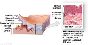
The epidermis is composed primarily of keratinocytes and is organized into distinct layers (strata). It is classified as either thin or thick skin based on the number of keratinocyte layers.
Keratinocytes: Most abundant cell type; produce keratin for waterproofing and protection.
Thin skin: Four layers; covers most of the body.
Thick skin: Five layers; found on palms and soles.
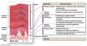
Layers (Strata) of the Epidermis
From deep to superficial, the layers of thick skin are:
Stratum basale (germinativum): Single layer of basal cells attached to the basement membrane; contains stem cells, melanocytes, and tactile (Merkel) cells.
Stratum spinosum: 8–10 layers of keratinocytes; contains dendritic (Langerhans) cells for immune defense.
Stratum granulosum: 3–5 layers; cells produce keratin and keratohyalin, then die.
Stratum lucidum: Only in thick skin; clear, thin layer of dead keratinocytes.
Stratum corneum: 15–30 layers of dead, keratinized cells; provides a water-resistant barrier.

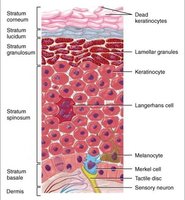
Specialized Cells of the Epidermis
Melanocytes: Produce melanin pigment for UV protection.
Merkel cells: Sensory receptors for touch, found in hairless skin.
Langerhans (dendritic) cells: Immune cells in the stratum spinosum.
Keratinization and Cell Turnover
Keratinization is the process by which keratinocytes fill with keratin and move from the stratum basale to the stratum corneum, where they are eventually shed. This process takes 7–10 days, and cells remain in the stratum corneum for about two weeks before being sloughed off.
Perspiration and Water Loss
Insensible perspiration: Water loss by diffusion through the stratum corneum (about 500 mL/day).
Sensible perspiration: Water excreted by sweat glands.
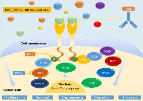
Epidermal Growth Factor (EGF)
EGF is a peptide growth factor produced by salivary glands and the duodenum. It promotes division of basal cells, accelerates keratin production, stimulates epidermal repair, and glandular secretion. EGF signaling involves several intracellular pathways, including PI3K/AKT, STAT, and MAPK cascades, which regulate cell proliferation, survival, adhesion, migration, and angiogenesis.
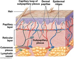
Dermis
Structure and Layers of the Dermis
The dermis is located between the epidermis and the subcutaneous layer. It anchors accessory structures and provides mechanical strength and elasticity to the skin. The dermis consists of two layers:
Papillary layer: Superficial areolar tissue; contains capillaries, lymphatics, and sensory neurons; forms dermal papillae.
Reticular layer: Dense irregular connective tissue; contains collagen and elastic fibers, blood vessels, and nerves.


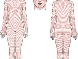
Dermal Strength, Elasticity, and Tension Lines
Collagen fibers: Provide tensile strength and resist stretching.
Elastic fibers: Allow stretching and recoil, contributing to skin flexibility (turgor).
Tension lines (cleavage lines): Patterns of collagen and elastic fibers; incisions parallel to these lines heal better.
Dermal Blood Supply and Innervation
Cutaneous plexus: Deep network of arteries in the reticular layer.
Subpapillary plexus: Network of small arteries in the papillary layer.
Innervation: Nerve fibers control blood flow, gland secretion, and monitor sensory receptors (e.g., Meissner and Pacinian corpuscles).
Subcutaneous Layer (Hypodermis)
The subcutaneous layer lies deep to the dermis and is composed primarily of adipose tissue. It stabilizes the position of the skin, stores energy, and provides insulation. Large arteries and veins are present in the superficial region, and this layer is a common site for subcutaneous injections.
Skin Color
Pigments Influencing Skin Color
Melanin: Produced by melanocytes; protects against UV radiation; more melanosomes in darker skin.
Carotene: Orange-yellow pigment from diet; can be converted to vitamin A.
Hemoglobin: Oxygenated blood gives skin a reddish hue; deoxygenated blood causes cyanosis (bluish color).
Clinical Correlations
Jaundice: Yellowing due to bile accumulation.
Vitiligo: Loss of melanocytes and skin color.
Addison’s disease and pituitary tumors: Can increase melanin production.
Accessory Structures
Hair and Hair Follicles
Hair is an accessory structure derived from the epidermis but located in the dermis. It serves protective, sensory, and thermoregulatory functions. Hair follicles produce hair and are associated with sebaceous glands and arrector pili muscles.
Regions: Hair root (anchored in skin), hair shaft (exposed above surface).
Layers: Medulla (core), cortex (middle), cuticle (surface), internal and external root sheaths, glassy membrane.
Types: Terminal hairs (thick, pigmented), vellus hairs (fine, unpigmented).
Hair color: Determined by melanocyte activity and genetics.
Exocrine Glands
Sebaceous glands: Secrete sebum (oil) into hair follicles or directly onto skin; lubricates and protects.
Apocrine sweat glands: Found in armpits, nipples, pubic region; secrete into hair follicles; produce odorous secretions.
Eccrine (merocrine) sweat glands: Widely distributed; secrete directly onto skin; important for thermoregulation.
Other glands: Mammary glands (milk), ceruminous glands (earwax).
Nails
Nails are protective structures composed of keratinized cells. They protect the tips of fingers and toes and can reflect underlying health conditions.
Nail body: Visible portion covering the nail bed.
Nail root: Site of nail production.
Lunula: Pale crescent near the root.
Repair and Aging of the Integument
Repair Process
Bleeding, inflammation, and scab formation occur after injury.
Macrophages clean debris; fibroblasts produce granulation tissue.
Scar tissue forms as healing progresses; keloids may develop.
Aging Effects
Epidermis thins, increasing infection risk.
Decreased dendritic cells, vitamin D3 production, melanocyte, and glandular activity.
Reduced blood supply, hair follicle function, and repair rate.
Dermis thins; elastic fibers decrease; fat distribution changes.
