 Back
BackThe Integumentary System: Structure, Function, and Clinical Relevance
Study Guide - Smart Notes
 Tailored notes based on your materials, expanded with key definitions, examples, and context.
The Integumentary System
Overview and Functions
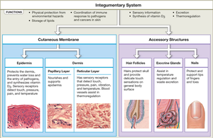
The integumentary system is the body's largest and most accessible organ system, comprising about 16% of total body weight and covering 1.5–2 m2. It serves as the first line of defense against environmental hazards and is essential for protection, excretion, thermoregulation, vitamin D3 synthesis, lipid storage, sensory detection, and immune coordination.
Protection: Shields underlying tissues from impact, abrasion, fluid loss, and chemical attack.
Excretion: Removes salts, water, and organic wastes via glands.
Temperature Maintenance: Regulates body temperature through insulation and evaporative cooling.
Vitamin D3 Synthesis: Produces cholecalciferol for calcium metabolism.
Lipid Storage: Stores lipids in dermal adipocytes and subcutaneous fat.
Sensory Detection: Monitors touch, pressure, pain, vibration, and temperature.
Immune Response Coordination: Responds to pathogens and cancers in the skin.
Structure of the Integumentary System
Cutaneous Membrane
The cutaneous membrane consists of two main layers: the epidermis and the dermis. Beneath the dermis lies the subcutaneous layer (hypodermis), which stabilizes the skin and stores fat.
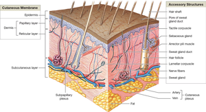
Epidermis
The epidermis is a stratified squamous epithelium providing physical protection, preventing water loss, and keeping microorganisms out. It is avascular, relying on diffusion from the dermis for nutrients.
Keratinocytes: Most abundant cells, produce keratin for strength and water resistance.
Strata: Layers include stratum basale, spinosum, granulosum, lucidum (in thick skin), and corneum.
Specialized Cells: Melanocytes (pigment production), dendritic cells (immune response), tactile cells (touch sensation).
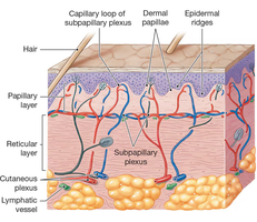
Dermis
The dermis lies beneath the epidermis and is divided into two layers:
Papillary Layer: Areolar tissue with capillaries, lymphatics, and sensory nerves.
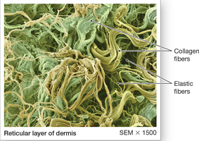
Reticular Layer: Dense irregular connective tissue with collagen and elastic fibers, providing strength and elasticity.
Subcutaneous Layer (Hypodermis)
The subcutaneous layer is composed mainly of adipose tissue, providing insulation, energy storage, and shock absorption. It contains large blood vessels and is a common site for drug injections.
Accessory Structures
Accessory structures include hair follicles, exocrine glands, and nails, all derived from the epidermis but located in the dermis or hypodermis.
Hair Follicles: Produce hair for protection, sensory input, and insulation.
Exocrine Glands: Sebaceous glands secrete sebum for lubrication; sweat glands (apocrine and eccrine) regulate temperature and excrete wastes.
Nails: Protect and reinforce the tips of fingers and toes.
Skin Pigmentation and Clinical Implications
Pigments
Melanin: Produced by melanocytes, protects against UV radiation. Two types: pheomelanin (red-yellow) and eumelanin (brown-black).
Carotene: Orange-yellow pigment, accumulates in epidermal cells and fat, can be converted to vitamin A.
Hemoglobin: Blood pigment, gives skin a reddish tint when oxygenated; cyanosis occurs when oxygen is low.
Clinical Conditions
Albinism: Lack of melanin production.
Vitiligo: Loss of melanocytes, resulting in white patches.
Jaundice: Yellowing due to liver dysfunction.
Addison's Disease: Increased ACTH causes skin darkening.
Vitamin D3 Synthesis
Process and Importance
UV radiation stimulates epidermal cells to produce cholecalciferol (vitamin D3), which is converted by the liver and kidneys to calcitriol. Calcitriol is essential for calcium and phosphate absorption in the intestine.
Deficiency: Leads to rickets, a disease of impaired bone development.
Hair and Hair Follicles
Structure and Function
Hair is produced by follicles and consists of a root and shaft. The hair bulb surrounds the papilla, and the matrix produces new hair cells. Hair types include lanugo (embryonic), vellus (fine), and terminal (thick).
Arrector Pili Muscle: Contracts to make hair stand erect (goosebumps).
Growth Cycle: Hair grows for 2–5 years, then is shed.
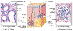
Exocrine Glands
Sebaceous and Sweat Glands
Sebaceous glands secrete sebum for lubrication and protection. Sweat glands are of two types:
Apocrine: Secrete odorous fluid into hair follicles; active at puberty.
Eccrine (Merocrine): Widely distributed, produce watery sweat for cooling and excretion.
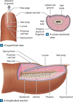
Nails
Structure and Function
Nails protect the dorsal surfaces of fingers and toes. The nail body covers the nail bed, and production occurs at the nail root. The cuticle (eponychium) and lunula are visible features.
Skin Repair and Healing
Phases of Repair
Inflammation Phase: Bleeding and inflammatory response.
Migration Phase: Formation of a scab and granulation tissue.
Proliferation Phase: Fibroblasts produce collagen, scab undermined.
Scarring Phase: Formation of inflexible scar tissue (keloids may develop).
Burns and Clinical Assessment
Types of Burns
First-degree: Damage to epidermis only.
Second-degree: Damage to epidermis and part of dermis; blistering.
Third-degree: Full-thickness burn; destroys epidermis, dermis, and hypodermis.
Rule of Nines
Used to estimate burn area by dividing the body into regions representing 9% of total surface area.
Aging and the Integumentary System
Effects of Aging
Epidermis thins, increasing risk of injury.
Dendritic cell numbers decrease, reducing immune response.
Vitamin D3 production declines, affecting bone health.
Melanocyte activity decreases, increasing sun sensitivity.
Glandular activity declines, causing dry skin.
Blood supply reduces, impairing thermoregulation.
Hair follicles become less active, leading to thinner, grayer hair.
Skin repair slows, increasing infection risk.
Summary Table: Layers and Functions of the Integumentary System
Layer/Structure | Main Components | Functions |
|---|---|---|
Epidermis | Keratinocytes, melanocytes, dendritic cells, tactile cells | Protection, water resistance, UV protection, sensory input |
Dermis | Papillary (areolar tissue), reticular (dense connective tissue) | Strength, elasticity, blood supply, sensory receptors |
Subcutaneous Layer | Adipose tissue, blood vessels | Insulation, energy storage, shock absorption |
Accessory Structures | Hair follicles, glands, nails | Protection, secretion, sensory input |
Key Equations
Vitamin D3 Synthesis:
