 Back
BackThe Integumentary System: Structure, Function, and Clinical Aspects
Study Guide - Smart Notes
 Tailored notes based on your materials, expanded with key definitions, examples, and context.
The Integumentary System
Overview and General Structure
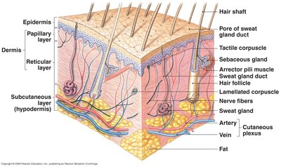
The integumentary system is the largest organ system in the human body, comprising approximately 16% of body weight and covering 1.5 to 2 square meters. It consists of the cutaneous membrane (skin), subcutaneous layer, and accessory structures such as hair follicles, nails, and exocrine glands. The skin is organized into three main layers: the epidermis, dermis, and subcutaneous (hypodermis) layer.
Cutaneous membrane: Includes the epidermis (outer epithelial layer) and dermis (connective tissue layer).
Subcutaneous layer (hypodermis): Connects skin to underlying tissues, contains adipose tissue for insulation and energy storage.
Accessory structures: Hair follicles, nails, and exocrine glands (sweat and oil glands).
Functions of the Integumentary System
Protection: Acts as a barrier against mechanical injury, pathogens, and water loss.
Temperature regulation: Blood vessels and sweat glands help regulate body temperature.
Nutrient storage: Stores lipids for energy and hormone synthesis.
Vitamin D3 synthesis: Initiates synthesis of vitamin D3 when exposed to UVB radiation.
Sensory detection: Contains receptors for touch, pain, temperature, and pressure.
Excretion: Removes waste products through sweat.
Skin Structure
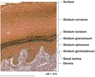
The Epidermis
The epidermis is a stratified squamous epithelium, primarily composed of keratinocytes. It is avascular and attached to the dermis by the basal lamina. Key cell types include keratinocytes (produce keratin), melanocytes (produce melanin), Langerhans cells (immune function), and Merkel cells (sensory function).
Keratinization: Process by which keratinocytes produce keratin and move from the basal layer to the surface, dying and forming the protective stratum corneum.
Layers of the epidermis (from deep to superficial):
Stratum basale (germinativum)
Stratum spinosum
Stratum granulosum
Stratum lucidum (only in thick skin)
Stratum corneum
Thin Skin vs. Thick Skin
Thin skin covers most of the body and has four layers of keratinocytes, while thick skin (found on palms and soles) has five layers, including a prominent stratum lucidum. Thick skin provides extra protection in areas subject to abrasion.
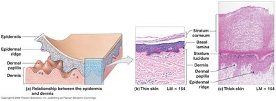
The Dermis
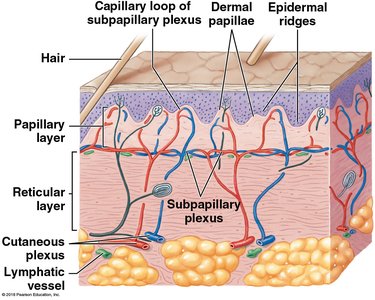
The dermis provides structural strength and elasticity to the skin. It is composed of two layers:
Papillary layer: Thin, superficial layer of loose areolar connective tissue with many capillaries and sensory receptors.
Reticular layer: Thick, deeper layer of dense irregular connective tissue, rich in collagen and elastin fibers.
The dermis contains blood vessels, nerves, and accessory structures such as hair follicles and glands.
The Subcutaneous Layer (Hypodermis)
The subcutaneous layer connects the skin to underlying tissues and organs. It consists of loose connective tissue and adipose tissue, providing insulation, energy storage, and cushioning. It also contains large blood vessels and nerves.
Physiological Functions of the Integumentary System
Nutrient Storage
The skin stores lipids in adipose tissue, which are used for energy production, synthesis of lipid hormones, and as components of cell membranes.
Temperature Regulation
Blood vessels in the dermis dilate to release heat and constrict to conserve heat. Sweat glands secrete water, which evaporates to cool the skin. The subcutaneous fat layer acts as insulation.
Excretion
Sweat glands excrete water, electrolytes (such as Na+ and Cl-), and organic wastes (urea, ammonia) from the body.
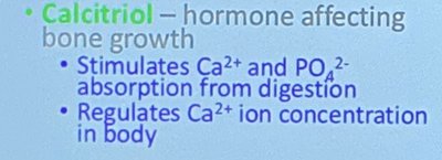
Vitamin D3 Synthesis
Keratinocytes exposed to UVB radiation synthesize cholecalciferol (vitamin D3) from cholesterol. Vitamin D3 is converted in the liver and kidneys to calcitriol, a hormone that regulates calcium and phosphate absorption and bone growth.
Key equation:
Sensory Detection
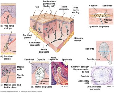
The skin contains various sensory receptors for detecting pain, temperature, touch, pressure, and vibration. These include free nerve endings, tactile (Meissner's) corpuscles, lamellated (Pacinian) corpuscles, and Merkel cells.
Protection
The integumentary system protects against mechanical abrasion, water loss, microbial invasion, and ultraviolet radiation. Melanocytes produce melanin, which absorbs UV radiation and reduces the risk of DNA mutations in skin cells.
Skin Color and Pigmentation
Determinants of Skin Color
Blood supply: Hemoglobin in red blood cells imparts a reddish hue; low oxygen levels cause cyanosis (bluish color).
Epidermal pigmentation: Beta-carotene (from diet) and melanin (produced by melanocytes) contribute to skin color.

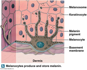
All humans have similar numbers of melanocytes; differences in skin color are due to the rate and amount of melanin production.
Disorders of the Epidermis
Common Disorders
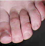
Warts: Caused by human papillomavirus (HPV).
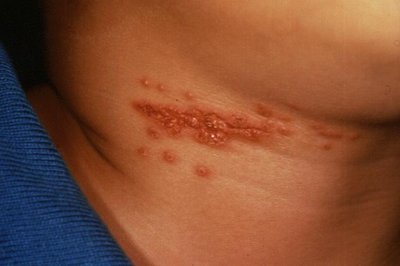
Herpes: Viral infections such as herpes simplex and varicella-zoster.
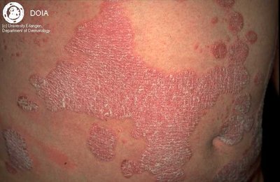
Psoriasis: Autoimmune disorder causing rapid skin cell turnover and scaling.
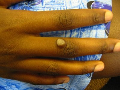
Corns and calluses: Localized hyperkeratosis due to friction or pressure.
Xerosis: Abnormal dryness of the skin.
Skin Cancer
Actinic keratosis: Precancerous lesion caused by UV exposure.
Basal cell carcinoma: Most common skin cancer, rarely metastasizes.
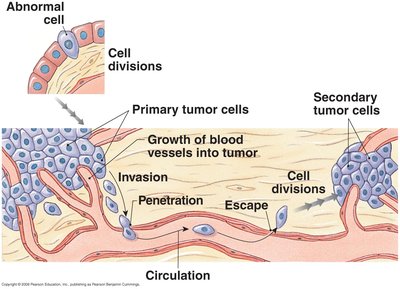
Squamous cell carcinoma: Second most common, can metastasize if untreated.
Neoplasms: Abnormal tissue growth due to genetic mutations; can be benign or malignant.
Carcinogens: Agents that cause cancer, such as UV light, chemicals, and viruses.
Accessory Structures of the Skin
Hair and Hair Follicles
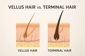
Hair is produced by hair follicles, which are invaginations of the epidermis into the dermis. Hair types include vellus (fine, unpigmented), terminal (coarse, pigmented), and club hair (no longer growing). Hair growth is cyclic and influenced by hormones and body chemistry.

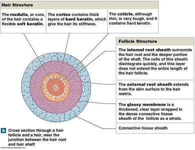
Hair structure: Medulla (soft keratin), cortex (hard keratin), cuticle (outer layer).
Follicle structure: Internal and external root sheaths, glassy membrane, connective tissue sheath.
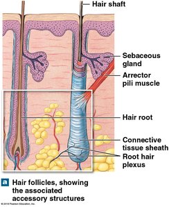
Arrector pili muscles attach to hair follicles and contract to raise hairs (goosebumps). Root hair plexuses are sensory nerve endings around hair follicles.
Exocrine Glands
Sebaceous glands: Secrete sebum (oil) onto hair shafts or skin surface; holocrine secretion.
Sudoriferous (sweat) glands: Merocrine (eccrine) glands produce watery sweat for cooling; apocrine glands secrete thicker fluid into hair follicles, active after puberty.
Ceruminous glands: Modified sweat glands in the ear canal, produce cerumen (earwax).
Mammary glands: Modified apocrine glands that produce milk.
Nails
Nails are protective coverings on the dorsal surfaces of fingers and toes. They are produced by the nail root (germinal matrix) and consist of dead, keratinized cells. Nail growth reflects overall body metabolism and health.
Wound Healing and Aging
Wound Healing
Skin repairs itself through a process involving inflammation, clot formation, cell migration, and tissue regeneration. Minimal scarring occurs if wound edges are well approximated and parallel to tension lines.
Burns and Ulcers
First-degree burns: Affect only the epidermis.
Second-degree burns: Damage epidermis and part of the dermis; blisters form.
Third-degree burns: Destroy epidermis, dermis, and subcutaneous tissue; require medical intervention.
Ulcers: Result from loss of circulation, leading to cell death and infection (e.g., bedsores, diabetic ulcers).
Aging and the Integumentary System
Epidermis and dermis thin
Decreased Langerhans cells (immune function)
Reduced blood flow and melanocyte activity
Decline in hair follicle function and glandular activity
Slower repair and reduced vitamin D production
Decreased elastin production, leading to wrinkles
