 Back
BackThe Integumentary System: Structure, Function, and Layers
Study Guide - Smart Notes
 Tailored notes based on your materials, expanded with key definitions, examples, and context.
The Integumentary System
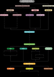
Introduction to the Integumentary System
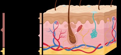
The integumentary system is an organ system consisting of the skin, hair, nails, glands, and sensory receptors. It is composed of several parts, including the cutaneous membrane (skin), which itself consists of the epidermis and dermis, and the subcutaneous layer (hypodermis) beneath the skin. The system serves multiple essential functions for the body.
Protection: Acts as a barrier against mechanical stresses, chemicals, UV light, and microbes.
Homeostasis: Maintains internal conditions, such as body temperature.
Sensation: Allows for sensory perception through nerve endings.
Communication: Facilitates expressive communication and emotions.
Vitamin D Synthesis: Skin synthesizes vitamin D when exposed to sunlight.
Excretion: Removes waste products via sweating.
Organization of the Integumentary System
The skin is organized into three main layers: the epidermis (outermost), dermis (middle), and hypodermis (deepest). Accessory structures include hair, nails, and glands. The epidermis is made of epithelial tissue, the dermis of connective tissue, and the hypodermis of adipose and areolar tissue.
Thermoregulation
Mechanisms of Thermoregulation
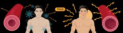
The integumentary system plays a crucial role in maintaining homeostasis, particularly through thermoregulation. This process involves two main methods:
Vasoconstriction & Vasodilation: Blood vessels in the dermis constrict (vasoconstriction) when cold, reducing heat loss, and dilate (vasodilation) when hot, increasing heat loss.
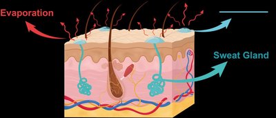
Sweating: Sweat glands secrete a water-based solution onto the skin, which cools the body as it evaporates.
These mechanisms are regulated by negative feedback loops to maintain stable internal body temperature.
The Epidermis
Cell Types in the Epidermis
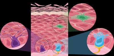
The epidermis is composed of stratified squamous epithelial tissue and contains four main cell types:
Keratinocytes: Most abundant; produce keratin, a tough, fibrous, water-resistant protein that provides mechanical strength and water resistance.
Melanocytes: Produce melanin, a pigment that protects skin from UV damage.
Dendritic Cells (Langerhans cells): Initiate immune responses and help prevent infection.
Tactile Epithelial Cells (Merkel cells): Specialized for touch sensation.
Keratinocyte Development and Function
Keratinocytes originate in the deepest layer of the epidermis and are pushed toward the surface, accumulating keratin and eventually dying. Tight junctions and desmosomes connect keratinocytes, forming a leak-proof barrier and providing mechanical resilience.
Keratin: Main component of hair and nails; provides water resistance and strength.
Barrier Function: Protects against water loss, pathogens, and harmful substances.

Layers of the Epidermis
The epidermis is organized into five distinct layers (strata), each with unique characteristics. The order from deep to superficial is:
Stratum Basale: Single row of proliferating cells; contains keratinocytes, melanocytes, and tactile epithelial cells.
Stratum Spinosum: Several rows of dividing keratinocytes; thickest layer in thin skin; contains dendritic cells.
Stratum Granulosum: Keratinocytes stop dividing, begin to harden and die; keratinization occurs; cells become waterproof.
Stratum Lucidum: Only in thick skin (palms, soles); flattened, dead, densely packed cells lacking organelles.
Stratum Corneum: Outermost layer; dead, keratin-filled cells; water-resistant glycolipid membranes; cells regularly shed.


Thin vs. Thick Skin
Skin varies in thickness depending on location:
Thin Skin: Covers most of the body; lacks stratum lucidum; contains hair follicles and oil glands; fewer sweat glands.
Thick Skin: Found on palms and soles; contains stratum lucidum; lacks hair follicles and oil glands; more sweat glands.
The Dermis
Structure and Layers of the Dermis
The dermis lies deep to the epidermis and consists of two layers:
Papillary Layer: Superficial; made of loose connective tissue; contains dermal papillae, blood vessels, lymphatic vessels, and Meissner corpuscles (touch receptors). Dermal papillae form epidermal ridges, enhancing grip and producing fingerprints.
Reticular Layer: Deep; made of dense irregular connective tissue; contains sweat and oil glands, hair roots, and Pacinian corpuscles (pressure receptors). Collagen and elastic fibers provide strength and flexibility; cleavage lines are parallel orientations of collagen fibers important for surgical healing.
The Hypodermis (Subcutaneous Layer)
Structure and Function of the Hypodermis
The hypodermis lies beneath the dermis and is not technically part of the skin. It is composed mostly of adipose tissue and some areolar connective tissue. The hypodermis anchors the skin to underlying tissues, acts as a shock absorber, and insulates the body to reduce heat loss.
Anchoring: Secures skin to muscles and bones.
Shock Absorption: Protects internal organs from mechanical injury.
Insulation: Helps maintain body temperature by reducing heat loss.
Energy Storage: Stores excess fat as an energy reserve.
Summary Table: Layers of the Skin
Layer | Main Tissue | Key Features |
|---|---|---|
Epidermis | Stratified squamous epithelium | Keratinocytes, melanocytes, dendritic cells, tactile epithelial cells; barrier function |
Dermis - Papillary Layer | Loose connective tissue | Dermal papillae, blood vessels, touch receptors |
Dermis - Reticular Layer | Dense irregular connective tissue | Collagen & elastic fibers, sweat/oil glands, hair roots, pressure receptors |
Hypodermis | Adipose & areolar tissue | Anchoring, shock absorption, insulation, energy storage |
Key Equations and Concepts
Negative Feedback Loop (Thermoregulation):
Evaporation Cooling:
