 Back
The Integumentary System: Structure, Function, and Clinical Relevance
Study Guide - Smart Notes
 Tailored notes based on your materials, expanded with key definitions, examples, and context.
The Integumentary System
Introduction to the Integumentary System
The integumentary system is a complex organ system that includes the skin, hair, nails, sweat glands, sebaceous glands, and mammary glands. It serves as the body's primary barrier against the external environment and plays critical roles in protection, sensation, thermoregulation, and metabolic functions.
Skin: The largest and most visible organ, providing a protective covering.
Accessory Structures: Includes hair, nails, and various glands that support the skin's functions.
Structure and Function of the Integumentary System
Major Components
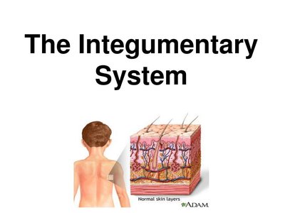
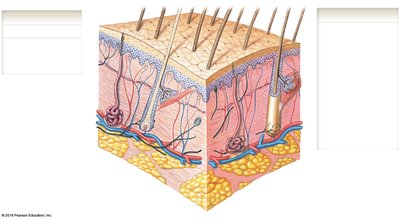
The integumentary system is organized into the cutaneous membrane (skin) and accessory structures. The cutaneous membrane consists of two main layers: the epidermis and the dermis. Deep to the dermis lies the hypodermis (subcutaneous layer), which is not technically part of the integument but supports its functions.
Epidermis: Superficial epithelial layer providing protection and sensory input.
Dermis: Underlying connective tissue layer containing blood vessels, nerves, and accessory structures.
Hypodermis: Subcutaneous tissue composed mainly of adipose tissue and major blood vessels.
Functions of the Integumentary System
Physical Protection: Shields underlying tissues from mechanical damage, pathogens, and chemicals.
Thermoregulation: Regulates body temperature through sweat production and blood flow.
Excretion: Removes metabolic wastes via sweat glands.
Synthesis: Produces vitamin D3 and stores lipids.
Sensation: Contains sensory receptors for touch, pressure, pain, and temperature.
Immune Defense: Coordinates immune responses to pathogens and cancers.
The Epidermis
Cell Types in the Epidermis
The epidermis is composed of four main cell types, each with specialized functions:
Keratinocytes: Produce keratin, a tough, protective protein.
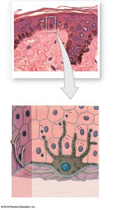
Melanocytes: Synthesize melanin, the pigment responsible for skin color and UV protection.
Merkel Cells: Sensory cells involved in touch perception.
Langerhans Cells: Immune cells that act as wandering macrophages.
Layers of the Epidermis
The epidermis is organized into distinct layers (strata), each with unique characteristics and functions:
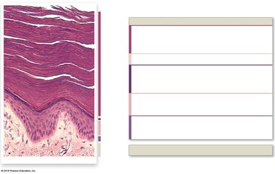
Stratum Basale: Deepest layer; contains stem cells, melanocytes, and Merkel cells. Site of active cell division.
Stratum Spinosum: Several layers of keratinocytes bound by desmosomes; contains Langerhans cells.
Stratum Granulosum: Keratinocytes produce keratin and keratohyalin; cells begin to die.
Stratum Lucidum: Present only in thick skin (palms, soles); cells are flat, lack organelles, and are densely packed with keratin.
Stratum Corneum: Most superficial layer; consists of dead, interlocking keratinized cells, providing a durable overcoat.
Thick vs. Thin Skin
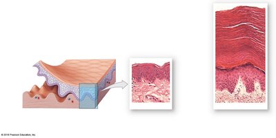
Thick Skin: Found on palms and soles; contains all five epidermal layers, including the stratum lucidum.
Thin Skin: Covers most of the body; lacks the stratum lucidum and has a thinner stratum corneum.
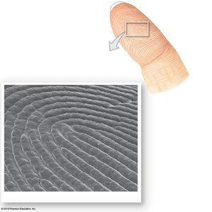
Dermal Ridges and Fingerprints
The stratum basale forms dermal ridges that extend into the dermis, creating fingerprints. These structures enhance grip and tactile sensitivity.
Determinants of Skin Color
Dermal Blood Supply: Reduced blood flow causes paleness; severe reduction leads to cyanosis (bluish skin).
Epidermal Pigments: Carotene (from diet) and melanin (produced by melanocytes) determine skin color. Melanin protects against UV radiation.
Genetic Control: Skin color is largely inherited but can be influenced by environmental factors such as UV exposure.
Vitamin D Synthesis
UV light converts a cholesterol-related precursor in the skin to vitamin D3 (cholecalciferol), which is then modified in the liver and kidneys to form the active hormone calcitriol, essential for calcium metabolism.
The Dermis
Dermal Organization
The dermis is divided into two layers:
Papillary Layer: Superficial; composed of loose connective tissue, contains dermal papillae, capillaries, and sensory neurons.
Reticular Layer: Deep; consists of dense irregular connective tissue, houses hair follicles, sweat glands, and sebaceous glands.
Wrinkles, Stretch Marks, and Tension Lines
Wrinkles: Result from aging, hormonal changes, and UV exposure, which reduce collagen and elastic fibers.
Stretch Marks: Occur when reticular fibers break due to rapid stretching (e.g., pregnancy, weight gain).
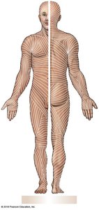
Tension Lines (Cleavage Lines): Collagen and elastic fibers align in parallel patterns; incisions made parallel to these lines heal with less scarring.
Blood and Nerve Supply
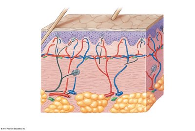
Blood Vessels: Form the cutaneous and subpapillary plexuses, essential for thermoregulation and nutrient delivery.
Nerve Fibers: Control blood flow, gland secretion, and monitor sensory receptors (e.g., tactile discs, Ruffini corpuscles, lamellated corpuscles).
The Subcutaneous Layer (Hypodermis)
Structure and Function
The subcutaneous layer lies deep to the dermis and is composed mainly of adipose tissue and major blood vessels. It stabilizes the skin, provides insulation, and serves as an energy reserve. It is the site for subcutaneous injections (e.g., hypodermic needles).
Accessory Structures
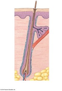
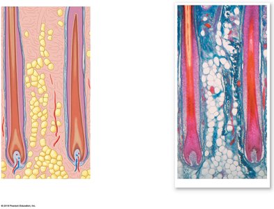
Hair Follicles and Hair
Hair is found almost everywhere on the body except the palms, soles, sides of fingers and toes, lips, and certain genital areas. Hair consists of the hair papilla, bulb, matrix, medulla, cortex, cuticle, root, and shaft.
Functions: Protection from UV light, insulation, sensory input, and prevention of foreign particle entry.
Types of Hair: Lanugo (embryonic), vellus (body hair), terminal (scalp, eyebrows, eyelashes).
Hair Color: Determined by melanin production; genetics, hormones, and age influence color.
Growth Cycle: Alternates between active growth and resting phases; male pattern baldness is hormonally influenced.
Glands in the Skin
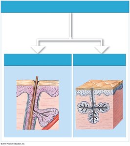
Sebaceous Glands: Secrete sebum to lubricate skin and hair; blockage can cause acne or folliculitis.
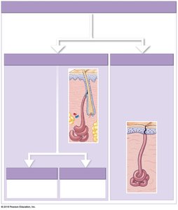
Sweat Glands:
Apocrine: Found in armpits and nipples; produce odorous secretions, may contain pheromones.
Eccrine (Merocrine): Widely distributed; produce watery sweat for cooling (sensible perspiration).
Ceruminous: Specialized apocrine glands in the ear canal; produce earwax (cerumen).
Mammary: Specialized apocrine glands for milk production.
Nails
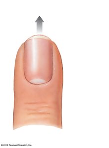
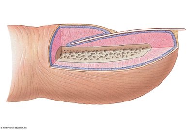
Nails protect the tips of fingers and toes. The nail body covers the nail bed, and the root is embedded near the bone. Other structures include the eponychium, hyponychium, lunula, and proximal nail fold.
Local Control and Aging of the Integumentary System
Local Control
Skin can respond to local influences by forming calluses or scar tissue.
Aging Effects
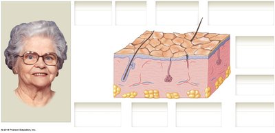
Epidermis and dermis become thinner.
Decrease in Langerhans cells, vitamin D production, melanocyte activity, and glandular activity.
Hair follicles stop functioning; skin repair slows; blood supply decreases, making heat dissipation difficult.
Changes in fat and hair distribution due to reduced sex hormones.
Summary Table: Layers and Structures of the Integumentary System
Layer/Structure | Main Components | Functions |
|---|---|---|
Epidermis | Keratinocytes, melanocytes, Merkel cells, Langerhans cells | Protection, sensation, vitamin D synthesis |
Dermis (Papillary) | Loose connective tissue, capillaries, sensory neurons | Support, nourishment, sensory input |
Dermis (Reticular) | Dense irregular connective tissue, glands, hair follicles | Strength, elasticity, housing accessory structures |
Hypodermis | Adipose tissue, major blood vessels | Insulation, energy storage, stabilization |
Accessory Structures | Hair, nails, glands | Protection, sensation, secretion |
