 Back
The Integumentary System: Structure, Function, and Clinical Relevance
Study Guide - Smart Notes
 Tailored notes based on your materials, expanded with key definitions, examples, and context.
The Integumentary System
Introduction
The integumentary system is the body's largest organ system, comprising the skin and its accessory structures. It plays a vital role in protection, sensation, thermoregulation, and homeostasis. This chapter explores the structure, function, and clinical significance of the integumentary system.
Skin Structure
Overview of Skin Layers
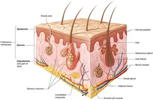
Cutaneous membrane (skin) consists of two main layers:
Epidermis: The superficial layer, composed of keratinized stratified squamous epithelium resting on a basement membrane. It is avascular and relies on diffusion from the dermis for nutrients and oxygen.
Dermis: Located deep to the epidermis, composed of loose connective tissue and dense irregular connective tissue. It is highly vascular and contains sensory receptors, glands, and hair follicles.
Hypodermis (subcutaneous layer): Not part of the skin but anchors it to underlying structures. Composed of loose connective and adipose tissue, it provides insulation and energy storage.
Functions of the Integumentary System
Protection
Mechanical trauma: The keratinized epithelium provides a durable, flexible barrier against physical injury.
Pathogen defense: The skin acts as a barrier to microorganisms and contains immune cells (e.g., dendritic cells) that destroy pathogens.
Environmental protection: Melanin absorbs UV radiation, and sebaceous secretions create an acidic environment (acid mantle) that inhibits microbial growth.
Water and electrolyte balance: Lipid-based secretions prevent excessive water loss or entry.
Sensation
The skin contains numerous sensory receptors that detect changes in temperature, pressure, pain, and touch, enabling the body to respond to environmental stimuli.
Thermoregulation
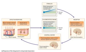
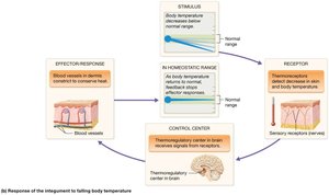
Thermoregulation is maintained through negative feedback loops involving the hypothalamus, sweat glands, and blood vessels.
When body temperature rises: Thermoreceptors signal the hypothalamus, which stimulates sweat production and cutaneous vasodilation, increasing heat loss.
When body temperature falls: Vasoconstriction reduces blood flow to the skin, conserving heat.
Excretion
The skin excretes small amounts of waste products such as urea and lactic acid through sweat.
Vitamin D Synthesis
UV radiation converts a cholesterol derivative in the epidermis to cholecalciferol (vitamin D3), which is then modified by the liver and kidneys to form calcitriol, essential for calcium absorption.
The Epidermis
Cell Types
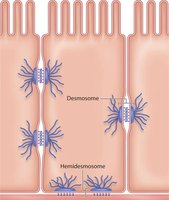
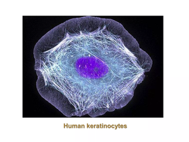
Keratinocytes: Make up 95% of the epidermis; produce keratin for strength and waterproofing; connected by desmosomes.
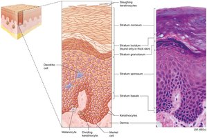
Dendritic (Langerhans) cells: Immune cells in the stratum spinosum.
Merkel cells: Sensory receptors for light touch, found in the stratum basale.
Melanocytes: Produce melanin pigment in the stratum basale.
Epidermal Layers (Strata)
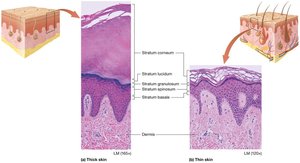
Stratum basale: Single layer of stem cells; site of mitosis and vitamin D synthesis.
Stratum spinosum: Thickest layer; metabolically active; contains dendritic cells.
Stratum granulosum: Contains granules of keratin and lipids; provides waterproofing.
Stratum lucidum: Present only in thick skin; clear, dead keratinocytes.
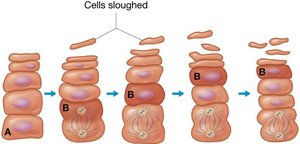
Stratum corneum: Outermost layer; dead, flattened keratinocytes; continuously shed.
Keratinocyte Life Cycle
Keratinocytes originate in the stratum basale, migrate upward, undergo keratinization, and are eventually sloughed off from the stratum corneum. This process takes 40–50 days.
Thick and Thin Skin
Differences
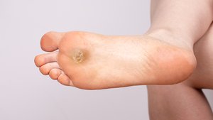
Thick skin: Found on palms and soles; contains all five strata; thick stratum corneum; no hair follicles.
Thin skin: Covers most of the body; lacks stratum lucidum; thinner layers; contains hair follicles and sebaceous glands.
Callus: Localized thickening of the stratum corneum due to repeated friction.
The Dermis
Structure and Function
Provides blood supply, anchors the epidermis, and contains sensory receptors.
Composed of two layers:
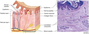
Papillary layer: Loose connective tissue; contains dermal papillae with capillaries and tactile (Meissner) corpuscles for light touch.
Reticular layer: Dense irregular connective tissue; contains collagen and elastic fibers, lamellated (Pacinian) corpuscles for pressure/vibration, glands, and hair follicles.
Skin Markings
Types and Clinical Relevance
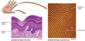
Dermal ridges: Form fingerprints; enhance grip.
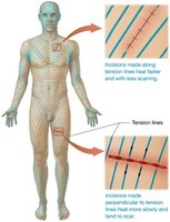
Tension (cleavage) lines: Indicate orientation of collagen bundles; important for surgical incisions.
Flexure lines: Deep creases near joints where skin is tightly bound to underlying structures.
Skin Pigmentation
Melanin
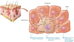
Produced by melanocytes; protects DNA from UV damage.
Transferred to keratinocytes via melanosomes; must be continually produced.
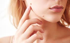
Variations include freckles (increased melanin production), moles (increased melanocyte number), and albinism (lack of melanin).

Carotene and Hemoglobin
Carotene: Yellow-orange pigment from diet; accumulates in stratum corneum.
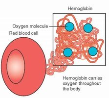
Hemoglobin: Oxygen-carrying protein in red blood cells; imparts a pinkish hue to skin depending on blood flow.
Skin Color as a Diagnostic Tool
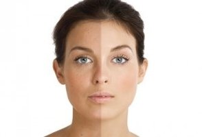
Erythema: Redness due to increased blood flow (e.g., exercise, fever).
Pallor: Paleness due to decreased blood flow (e.g., cold, shock).
Cyanosis: Bluish hue due to low oxygen saturation; indicates respiratory or circulatory issues.


Accessory Structures of the Integument
Hair
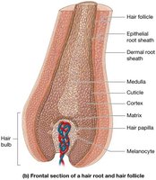
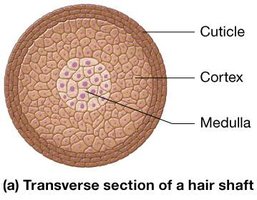
Composed of dead keratinized cells; found everywhere except thick skin, lips, and parts of external genitalia.
Functions: Protection (scalp, eyes, nose), sensation, minor role in thermoregulation.
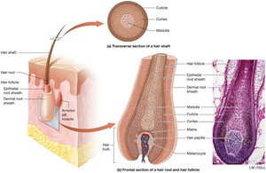
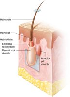
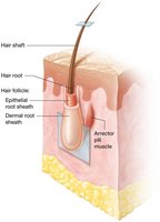
Structure: Shaft (above skin), root (in dermis), hair follicle, matrix (growth zone), and associated arrector pili muscle (causes piloerection/goosebumps).
Nails
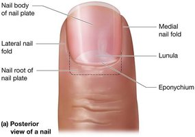
Hard structures at the ends of digits; composed of hard keratin.
Parts: Nail plate (body and root), nail matrix (growth zone), nail folds, and hyponychium.
Functions: Protection and precise manipulation of objects.
Glands
Sweat (sudoriferous) glands: Eccrine (thermoregulation), apocrine (odoriferous, active after puberty), ceruminous (ear wax), and mammary (milk production).
Sebaceous glands: Secrete sebum (oil) into hair follicles; lubricates and waterproofs skin and hair; antibacterial properties.
Burns
Classification and Clinical Relevance
First-degree: Only epidermis affected; redness and mild pain.
Second-degree: Epidermis and part/all of dermis; blistering and scarring.
Third-degree: Full thickness (epidermis, dermis, hypodermis, possibly deeper); not initially painful due to nerve destruction; high risk of dehydration and infection.
Rule of Nines: Used to estimate the percentage of body surface area affected by burns for clinical assessment and treatment planning.
Burn Degree | Layers Involved | Symptoms |
|---|---|---|
First-degree | Epidermis | Redness, mild pain |
Second-degree | Epidermis, part/all dermis | Pain, blistering, scarring |
Third-degree | All skin layers, possibly deeper | No initial pain, severe tissue loss, dehydration risk |
*Additional info: The "Rule of Nines" divides the body into 11 areas, each representing 9% of total body surface area, to quickly estimate burn extent in emergency settings.*
