 Back
The Integumentary System: Structure, Function, and Pathology
Study Guide - Smart Notes
 Tailored notes based on your materials, expanded with key definitions, examples, and context.
The Integumentary System
Overview and Skin Structure
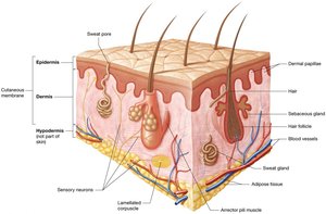
The integumentary system is the body's largest organ system, accounting for 10–15% of total body weight. It is essential for protection, homeostasis, and sensory perception. The skin, or cutaneous membrane, consists of two main components:
Epidermis: The superficial layer composed of keratinized stratified squamous epithelium resting on a basement membrane.
Dermis: Located beneath the epidermis and basement membrane, made of loose and dense irregular connective tissue.
Accessory structures embedded in the skin include sweat glands, sebaceous glands, hair, and nails. The hypodermis (superficial fascia or subcutaneous fat) lies deep to the dermis, anchoring the skin to deeper structures and providing insulation and energy storage.
Functions of the Integumentary System
The integumentary system performs several critical functions:
Protection: Provides a durable, flexible barrier against mechanical trauma, pathogens, and environmental hazards. The acid mantle inhibits pathogen growth, and immune cells destroy invaders.
Sensation: Sensory receptors in the skin detect changes in internal and external environments, including heat, cold, and pain.
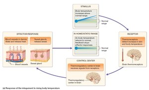
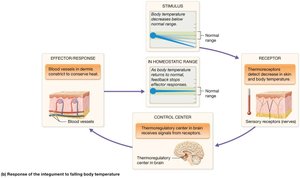
Thermoregulation: Maintains stable internal temperature via negative feedback loops. When body temperature rises, sweat glands and cutaneous vasodilation help cool the body. When temperature drops, vasoconstriction conserves heat.
Excretion: Eliminates waste products and toxins, supplementing the kidneys.
Vitamin D Synthesis: UV exposure converts a cholesterol precursor to cholecalciferol, which is modified to calcitriol, essential for calcium absorption.
The Epidermis
Cell Types and Layers
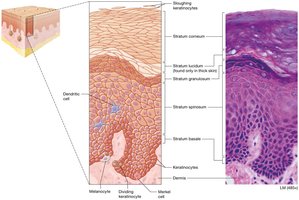
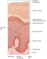
The epidermis is the most superficial skin layer, composed primarily of keratinocytes (about 95%), which produce keratin for strength and protection. Other cell types include:
Dendritic (Langerhans) cells: Immune phagocytes in the stratum spinosum.
Merkel cells: Sensory receptors for light touch in the stratum basale.
Melanocytes: Produce melanin pigment in the stratum basale.
The epidermis consists of five distinct strata (layers), from deep to superficial:
Stratum basale: Single layer of stem cells, most metabolically active.
Stratum spinosum: Thickest layer, still metabolically active.
Stratum granulosum: Contains keratin bundles and lipid-based granules for waterproofing.
Stratum lucidum: Clear, dead keratinocytes; only in thick skin.
Stratum corneum: Outermost layer of dead, flattened keratinocytes.
Keratinocyte Life Cycle
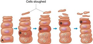
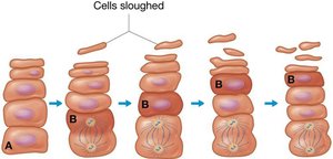
Keratinocytes originate in the stratum basale and migrate through each layer, eventually being shed from the stratum corneum. This process takes 40–50 days.
Thick and Thin Skin
Skin varies in thickness depending on location and function:
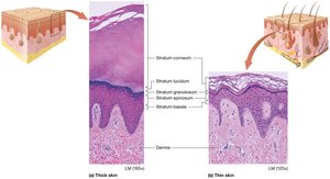
Thick skin: Found on palms and soles; contains all five strata, thick stratum corneum, no hair follicles, many sweat glands.
Thin skin: Covers most of the body; only four layers (no stratum lucidum), thinner strata, numerous hairs, sweat, and sebaceous glands.
Callus: Additional stratum corneum layers formed in response to repetitive pressure.
The Dermis
Structure and Layers
The dermis is a highly vascular layer beneath the epidermis, providing blood supply, sensory receptors, and anchoring the epidermis. It consists of two layers:
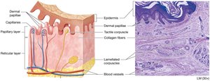
Papillary layer: Thinner, superficial layer of areolar loose connective tissue. Contains dermal papillae with capillaries and tactile (Meissner) corpuscles for light touch.
Reticular layer: Deep, thicker layer of dense irregular connective tissue. Contains collagen bundles for strength, elastic fibers for flexibility, lamellated (Pacinian) corpuscles for pressure and vibration, blood vessels, glands, and adipose tissue.
Skin Markings
Skin markings arise from interactions between the dermis and epidermis:
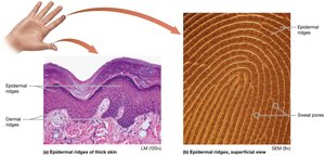
Dermal ridges: Prominent areas of thick collagen bundles, creating epidermal ridges (fingerprints) for enhanced grip.
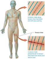
Tension (cleavage) lines: Indentations from gaps between collagen bundles, important for surgical incisions.
Flexure lines: Deep creases where the reticular layer is tightly anchored to deeper structures.
Skin Pigmentation
Melanin
Skin color is primarily determined by melanin, produced by melanocytes in the stratum basale. Melanin protects keratinocyte DNA from UV-induced mutations and must be produced continuously. Its synthesis increases with UV exposure, leading to tanning.
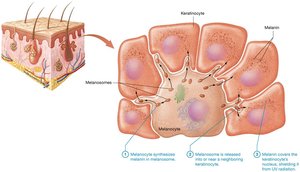
Immediate tanning: Oxidation of existing melanin.
Delayed tanning: Increased melanin production after DNA damage.
Secondary function: Reduces vitamin D synthesis, affecting calcium homeostasis.
Skin color differences are due to tyrosinase activity and melanin type, not melanocyte number. Common variations include freckles, moles, and albinism.
Carotene and Hemoglobin
Carotene: Yellow-orange pigment from diet, accumulates in stratum corneum.
Hemoglobin: Iron-containing protein in red blood cells, gives skin a pinkish hue based on blood flow.
Skin Color as a Diagnostic Tool
Erythema: Increased blood flow, red skin (exercise, trauma, fever).
Pallor: Decreased blood flow, pale skin (cold, stress).
Cyanosis: Low oxygen, bluish skin (respiratory or circulatory issues).
Accessory Structures: Hair, Nails, and Glands
Hair
Hair (pili) is a filamentous structure derived from epithelium, found almost everywhere except thick skin, lips, and parts of external genitalia. It provides protection, sensory input, and limited thermoregulation.
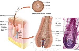
Hair Structure
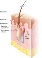
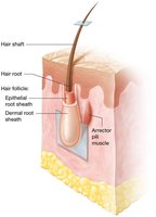
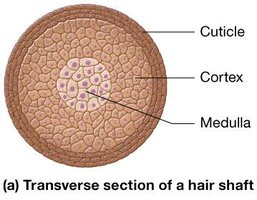
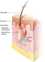
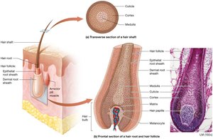
Shaft: Visible portion, composed of dead, keratinized cells.
Root: Embedded in dermis, surrounded by sensory neurons and the hair follicle.
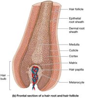
Matrix: Keratinocytes at the base of the root, actively dividing.
Root sheath: Outer component anchors follicle; inner component tightly attached to hair root.
Medulla, cortex, cuticle: Three regions in transverse section; medulla (soft keratin), cortex (hard keratin), cuticle (hard keratin, mechanical strength).
Dermal root sheath: Connective tissue support.
Arrector pili muscles: Smooth muscle bands causing piloerection (goosebumps).
Hair Pigment and Texture
Lanugo: Thin, nonpigmented fetal hair.
Terminal hair: Thick, pigmented hair (scalp, eyebrows).
Vellus hair: Thin, nonpigmented hair (body).
Hair color: Determined by melanin; blond (little melanin), black (lots), red (special pigment with iron). Hair turns gray/white with age.
Nails
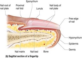
Nails are hard structures at the ends of digits, composed of stratified squamous epithelium filled with hard keratin. They protect underlying tissue and aid in gripping.
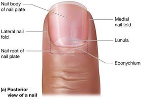
Nail plate: Visible component, divided into nail body and nail root.
Proximal nail fold: Covers nail root; eponychium (cuticle).
Medial/lateral nail folds: Reinforce nail plate.
Hyponychium: Stratum corneum attaching free edge to nail bed.
Lunula: Half-moon region of nail plate, accumulation of keratin.
Nail matrix: Site of nail growth.
Glands
Skin contains two main types of glands:
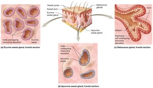
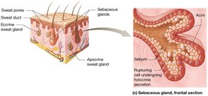
Sweat (sudoriferous) glands: Produce sweat; four types:
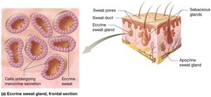
Eccrine: Most prevalent, simple coiled tubular glands, secrete watery sweat.
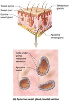
Apocrine: Large glands in specific regions, secrete protein-rich fluid into hair follicles.
Ceruminous: Modified apocrine glands, produce cerumen (ear wax).
Mammary: Specialized sweat glands, produce milk.
Sebaceous glands: Branched glands producing oily sebum, released by holocrine secretion. Sebum coats hair, inhibits water loss, and kills bacteria.
Pathology of the Skin
Acne
Acne vulgaris is a common skin disorder caused by accumulation of sebum and dead cells in sebaceous glands, leading to comedones (blackheads) and sometimes infection and inflammation (pustules).
Wounds and Burns
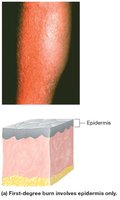
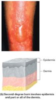
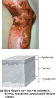
Wounds disrupt skin integrity and include lacerations, burns, and skin cancers. Burns are classified by depth:
First-degree: Only epidermis; erythema and mild pain.
Second-degree: Epidermis and part/all of dermis; pain, blistering, scarring.
Third-degree: Epidermis, dermis, hypodermis, possibly deeper tissues; major tissue damage, scarring, risk of dehydration and infection.
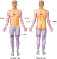
The rule of nines estimates burn extent by dividing the body into areas representing 9% of total body area.
Skin Cancer
Skin cancer is caused by DNA mutations leading to uncontrolled cell division. Three main types:
Basal cell carcinoma: Most common, arises from keratinocytes in stratum basale, rarely metastasizes.
Squamous cell carcinoma: Second most common, arises from keratinocytes in stratum spinosum, more likely to metastasize.
Malignant melanoma: Cancer of melanocytes, highly metastatic, prognosis depends on early detection and tumor depth.
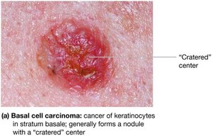
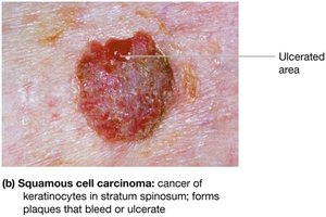
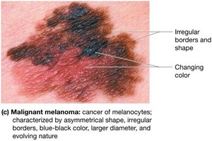
The ABCDE rule helps distinguish melanoma: Asymmetry, Border irregularity, Color variation, Diameter >6 mm, Evolving shape/size.
Summary Table: Layers of the Skin
Layer | Location | Main Features |
|---|---|---|
Epidermis | Superficial | Keratinized stratified squamous epithelium; avascular; five strata |
Dermis | Deep to epidermis | Loose and dense irregular connective tissue; vascular; papillary and reticular layers |
Hypodermis | Deep to dermis | Loose connective and adipose tissue; anchors skin; not part of skin |
Summary Table: Types of Skin Cancer
Type | Origin | Features | Metastasis |
|---|---|---|---|
Basal cell carcinoma | Stratum basale keratinocytes | Nodule with central crater | Rare |
Squamous cell carcinoma | Stratum spinosum keratinocytes | Scaly plaques, may ulcerate | Possible |
Malignant melanoma | Melanocytes | Irregular shape, color, evolving | High |
Key Equations
Vitamin D synthesis pathway:
UV exposure:
Liver modification:
Kidney modification:
Calcitriol is the active form of vitamin D, essential for calcium absorption.
Additional info: The notes have been expanded with academic context and tables for clarity and completeness.
