 Back
BackThe Integumentary System: Structure, Function, and Clinical Aspects
Study Guide - Smart Notes
 Tailored notes based on your materials, expanded with key definitions, examples, and context.
The Integumentary System
Overview
The integumentary system is a complex organ system that includes the skin and its associated structures. It serves as the body's primary barrier against the external environment and plays critical roles in protection, sensation, thermoregulation, and metabolic functions.
Components: Skin, sweat (sudoriferous) glands, oil (sebaceous) glands, hair, nails, and subcutaneous tissue.
Functions: Protection, temperature regulation, sensation, metabolic activity, blood reservoir, and excretion.
Structure of the Skin
Layers of the Skin
The skin consists of two main layers and an associated subcutaneous layer.
Epidermis: Outermost layer, composed of keratinized stratified squamous epithelium; avascular.
Dermis: Underlies the epidermis; composed mainly of dense connective tissue; vascular.
Subcutaneous Tissue (Hypodermis): Not part of the skin proper, but anchors skin to underlying structures; composed mostly of adipose tissue.
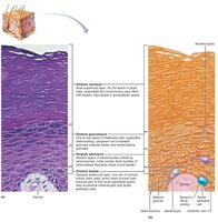
Epidermis: Cell Types and Layers
The epidermis contains four main cell types and is organized into distinct layers (strata).
Keratinocytes: Produce keratin, the protein that provides protective properties.
Melanocytes: Produce melanin pigment, which protects against UV damage.
Dendritic (Langerhans) Cells: Immune cells that patrol the epidermis.
Tactile (Merkel) Cells: Sensory receptors for touch.
Layers of the Epidermis:
Stratum basale: Deepest layer; mitotically active stem cells; contains melanocytes.
Stratum spinosum: Several layers thick; contains keratinocytes, melanosomes, and dendritic cells.
Stratum granulosum: Cells flatten, keratinization begins, water-resistant glycolipids accumulate.
Stratum lucidum: Present only in thick skin (palms, soles); clear, dead keratinocytes.
Stratum corneum: Most superficial; 20–30 rows of dead, keratinized cells; barrier function.
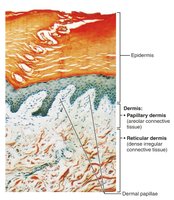
Dermis: Layers and Features
The dermis is a strong, flexible connective tissue layer with two distinct regions.
Papillary Dermis: Superficial; areolar connective tissue; contains dermal papillae with capillary loops, nerve endings, and touch receptors.
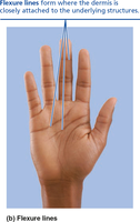
Reticular Dermis: Deep; dense irregular connective tissue; provides strength, elasticity, and hydration; contains cleavage lines and flexure lines.
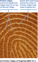
Dermal Modifications and Clinical Aspects
Friction ridges: Enhance grip and touch; form fingerprints.
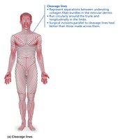
Cleavage (tension) lines: Parallel collagen bundles; important for surgical incisions.
Flexure lines: Dermal folds at joints; visible on hands and feet.
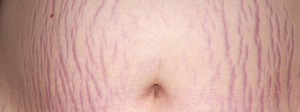
Striae (stretch marks): Dermal tears from extreme stretching.
Blisters: Fluid-filled pockets separating epidermal and dermal layers.
Pigments and Skin Color
Melanin, Carotene, and Hemoglobin
Skin color is determined by three main pigments.
Melanin: Produced by melanocytes; shields DNA from UV; more sun exposure increases melanin production.
Carotene: Yellow-orange pigment; accumulates in stratum corneum and subcutaneous tissue; can be converted to vitamin A.
Hemoglobin: Pinkish hue in fair skin due to transparency and underlying blood.
Clinical Significance:
Cyanosis: Bluish skin due to low oxygenation.
Pallor: Pale skin from stress, anemia, or low blood pressure.
Erythema: Redness from fever, inflammation, or allergy.
Jaundice: Yellow skin from liver disorders.
Bruises: Blood leakage under skin.
Hyperpigmentation: May indicate endocrine disorders.
Hair
Structure and Function
Hair is composed of dead, keratinized cells and serves various protective and sensory functions.
Regions: Root (within scalp, keratinization ongoing), shaft (extends above scalp, keratinization complete).
Parts of Hair Shaft: Medulla (central core), cortex (surrounds medulla), cuticle (outer layer).
Pigmentation: Melanocytes produce melanin; red hair has pheomelanin; gray/white hair results from decreased melanin and air bubbles.
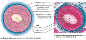
Hair Follicle Structure
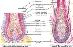
Hair follicle: Extends from epidermis to dermis; contains hair bulb and sensory nerve endings.
Wall: Peripheral connective tissue sheath, glassy membrane, epithelial root sheath.
Hair papilla: Supplies nutrients; if destroyed, hair growth ceases.
Hair matrix: Actively dividing cells producing hair.
Arrector pili: Smooth muscle causing "goose bumps."
Types and Growth of Hair
Vellus hair: Fine, pale body hair.
Terminal hair: Coarse, long hair (scalp, eyebrows, pubic, axillary regions).
Growth cycles: Active and resting phases; affected by nutrition and hormones.
Clinical Aspects:
Hirsutism: Excessive terminal hair in females due to androgen excess.
Baldness: Age-related hair loss; male pattern baldness is androgen-dependent.
Telogen effluvium: Abrupt hair thinning from stress or illness.
Nails
Structure and Function
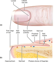
Nails are protective, scale-like modifications of the epidermis containing hard keratin.
Parts: Root (embedded), nail plate/body (visible), free edge.
Nail bed: Epidermis under nail plate.
Nail matrix: Growth region.
Nail folds: Skin folds overlapping nail border; cuticle (eponychium), hyponychium (under free edge).
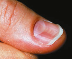
Lunule: White, thickened nail matrix.
Clinical Aspects
Yellow nails: May indicate respiratory or thyroid disorders.
Thickened yellow nails: Fungal infection.
Koilonychia (spoon nail): Iron deficiency.
Beau’s lines: Horizontal lines indicating severe illness.
Skin Appendages: Glands
Sweat Glands
Sweat glands regulate body temperature and excrete wastes.
Eccrine (merocrine) glands: Most numerous; secrete watery sweat for thermoregulation.
Apocrine glands: Located in axillary and anogenital areas; secrete viscous sweat; function may be related to scent.
Modified apocrine glands: Ceruminous (earwax) and mammary (milk) glands.
Oil (Sebaceous) Glands
Sebaceous glands: Secrete sebum (oil); bactericidal; softens skin and hair; most active after puberty.
Clinical Aspects:
Acne: Infectious inflammation of sebaceous glands.
Cradle cap (seborrhea): Overactive sebaceous glands in infants.
Functions of Skin
Protection
The skin acts as a barrier through chemical, physical, and biological mechanisms.
Chemical: Acid mantle, antimicrobial proteins, melanin.
Physical: Keratinized cells, glycolipids, limited permeability.
Biological: Dendritic cells (epidermis), macrophages (dermis).
Body Temperature Regulation
Insensible perspiration: Baseline sweat production.
Sensible perspiration: Increased sweat for cooling.
Cold response: Dermal blood vessels constrict to conserve heat.
Cutaneous Sensations
Touch: Meissner’s corpuscles, Merkel cells.
Pressure: Pacinian corpuscles.
Pain: Free nerve endings.
Hair movement: Hair follicle receptors.
Metabolic Functions
Vitamin D synthesis: Required for calcium absorption.
Activation of hormones: Keratinocytes convert cortisone to hydrocortisone.
Collagenase production: Aids in collagen turnover.
Blood Reservoir and Excretion
Blood reservoir: Skin holds up to 5% of blood volume.
Excretion: Removal of nitrogenous wastes, salt, and water via sweat.
Skin Disorders: Cancer and Burns
Skin Cancer
Basal cell carcinoma: Most common, least malignant; arises from stratum basale.
Squamous cell carcinoma: Second most common; arises from stratum spinosum; can metastasize.
Melanoma: Cancer of melanocytes; most dangerous, highly metastatic.
Detection:
ABCDE rule: Asymmetry, Border irregularity, Color variation, Diameter (>6 mm), Evolution.
Burns
First-degree: Epidermal damage only; redness, pain.
Second-degree: Epidermal and upper dermal damage; blisters.
Third-degree: Full-thickness; entire skin destroyed; requires grafting.
Rule of Nines: Used to estimate burn area and fluid loss.
Developmental Aspects
Embryonic Development
Epidermis: Develops from ectoderm.
Dermis and hypodermis: Develop from mesoderm.
Lanugo: Fine fetal hair; replaced by vellus hair before birth.
Infancy to Adulthood
Vernix caseosa: Protective substance from sebaceous glands.
Milia: Small white spots on newborn skin.
Acne: Increased gland activity during adolescence.
Aging Skin
Thinning: Reduced epidermal replacement.
Dryness and wrinkles: Decreased sebaceous activity and elasticity.
Increased cancer risk: Fewer melanocytes and dendritic cells.
Hair thinning: Common with age.
Prevention: UV protection is essential.
