 Back
Ch 5: The Integumentary System: Structure, Function, and Repair
Study Guide - Smart Notes
 Tailored notes based on your materials, expanded with key definitions, examples, and context.
Ch 5: The Integumentary System
Overview
The integumentary system is composed of the skin and its accessory structures, including hair, nails, and glands. It serves as the body's primary barrier against environmental hazards, regulates temperature, and provides sensory information.
The Epidermis
Structure and Function
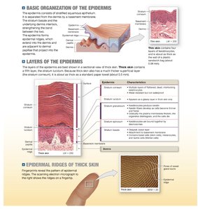
The epidermis is a stratified squamous epithelium that provides physical protection, prevents water loss, and helps keep microorganisms outside the body. It is avascular, relying on diffusion from the underlying dermis for nutrients and oxygen. The cells closest to the basement membrane are metabolically active, while the superficial cells are dead.
Keratinocytes: The most abundant cells, producing the tough protein keratin.
Melanocytes: Produce melanin, contributing to skin color and UV protection.
Tactile (Merkel) cells: Sensory cells for touch.
Dendritic (Langerhans) cells: Immune cells that defend against pathogens and cancer.
Layers of the Epidermis
The epidermis consists of several layers (strata), each with distinct functions:
Layer | Characteristics |
|---|---|
Stratum basale | Deepest layer; contains stem cells, melanocytes, and tactile cells; forms epidermal ridges. |
Stratum spinosum | 8–10 layers of keratinocytes; contains dendritic cells; some cell division occurs. |
Stratum granulosum | 3–5 layers; keratin and keratohyalin production; cells flatten and die. |
Stratum lucidum | Only in thick skin; clear, densely packed, keratin-filled cells. |
Stratum corneum | 15–30 layers of dead, keratinized cells; water-resistant barrier; cells are shed in sheets. |
Epidermal ridges (fingerprints) are unique and increase grip by enhancing friction.
Epidermal Growth Factor (EGF)
EGF is a peptide growth factor that stimulates epidermal cell division, keratin production, and repair. It is used in tissue culture and clinical treatments for burns.
The Dermis
Structure and Function
The dermis supports the epidermis and contains blood vessels, nerves, and accessory structures. It is divided into two layers:
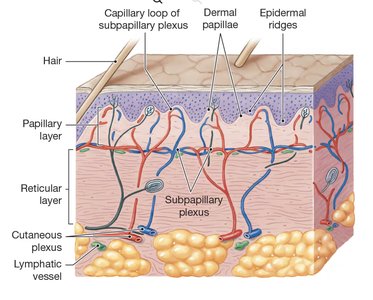
Papillary layer: Areolar tissue with capillaries, lymphatics, and sensory neurons; forms dermal papillae.
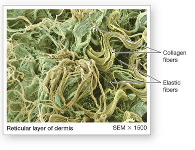
Reticular layer: Dense irregular connective tissue with collagen and elastic fibers, providing strength and elasticity.
Dermal Strength and Elasticity
Collagen fibers resist stretching, while elastic fibers allow flexibility. Skin turgor depends on water content. Aging, hormones, and UV exposure reduce elasticity, leading to wrinkles and stretch marks.
Tension Lines
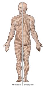
Tension (cleavage) lines are patterns of collagen and elastic fiber bundles. Surgical incisions parallel to these lines heal better and with less scarring.
Dermal Blood Supply
The dermis contains two vascular plexuses:
Cutaneous plexus: At the border of the reticular layer and subcutaneous layer.
Subpapillary plexus: Supplies the papillary layer and epidermis.
Trauma to dermal blood vessels can cause contusions (bruises).
Innervation of the Skin
Sensory receptors in the skin detect touch, pressure, pain, and temperature. Nerve fibers also regulate blood flow and glandular secretion.
The Subcutaneous Layer (Hypodermis)
Structure and Function
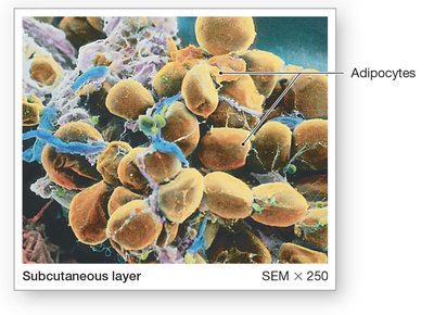
The subcutaneous layer (hypodermis) lies below the dermis and stabilizes the skin while allowing movement. It consists mainly of adipose tissue, serving as an energy reserve, insulator, and shock absorber. Distribution of subcutaneous fat varies by age, sex, and hormones.
Sunlight and Vitamin D3 Production
Interaction with the Integument
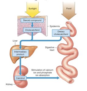
UV radiation stimulates epidermal cells to convert a steroid into cholecalciferol (vitamin D3). The liver and kidneys then convert it to calcitriol, which is essential for calcium and phosphate absorption in the intestine. Deficiency can lead to rickets in children.
Hair
Structure and Function
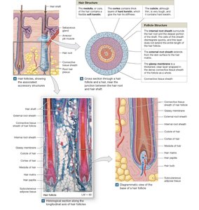
Hair is composed of keratinized dead cells produced in hair follicles. It protects, insulates, and serves as a sensory receptor. Hair structure includes the medulla, cortex, and cuticle, surrounded by follicle layers (internal root sheath, external root sheath, glassy membrane).
Hair Growth and Types
Growth cycle: Hair grows, then becomes inactive (club hair) and is shed.
Types: Lanugo (fetal), vellus (fine, unpigmented), terminal (thick, pigmented).
Color: Determined by melanin type and amount; decreases with age.
Glands of the Skin
Sebaceous (Oil) Glands
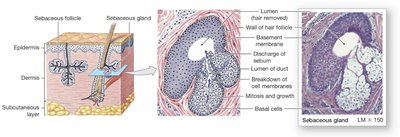
These holocrine glands secrete sebum into hair follicles or directly onto the skin. Sebum lubricates, protects, and inhibits bacterial growth. Overactivity can lead to seborrheic dermatitis (dandruff, cradle cap).
Sweat (Sudoriferous) Glands
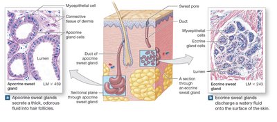
Apocrine glands: Found in armpits, nipples, and pubic region; secrete into hair follicles; produce odorous sweat; active at puberty.
Eccrine (merocrine) glands: Widely distributed; secrete directly onto skin; important for thermoregulation and excretion.
Other Glands
Mammary glands: Modified apocrine glands for milk production.
Ceruminous glands: Produce earwax (cerumen) in the external ear canal.
Nails
Structure and Function
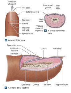
Nails are keratinized epidermal cells that protect the tips of fingers and toes and aid in manipulation. The nail body covers the nail bed, and growth occurs at the nail root. The eponychium (cuticle) and lunula are notable features.
Repair of the Integument
Phases of Skin Repair
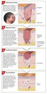
After injury, the skin repairs itself in four phases:
Inflammation phase: Bleeding and inflammation occur; mast cells trigger response.
Migration phase: Scab forms; cells migrate to repair the area; granulation tissue develops.
Proliferation phase: Fibroblasts produce collagen; tissue is rebuilt.
Scarring phase: Scar tissue forms; original tissue structure is not fully restored.
Excessive scar tissue may form keloids. Tattoos and other skin modifications involve the dermis and may result in permanent marks or scarring.
