 Back
The Integumentary System: Structure, Function, and Clinical Relevance
Study Guide - Smart Notes
 Tailored notes based on your materials, expanded with key definitions, examples, and context.
The Integumentary System
Overview and Components
The integumentary system is the body's largest organ system, serving as the primary interface between the body and the external environment. It consists of the skin, sweat and oil glands, hair, hair follicles, and nails. The skin alone covers 1.2–2.2 square meters, weighs 8–10 lbs, and varies in thickness from 1.5–4 mm.
Skin: The main protective barrier.
Sweat & Oil Glands: Involved in thermoregulation and lubrication.
Hair and Hair Follicles: Provide protection and sensory input.
Nails: Protect the distal phalanges and enhance fine touch.

Functions of the Integumentary System
The integumentary system performs several vital functions essential for homeostasis and survival:
Protection: Acts as a physical, chemical, and biological barrier against pathogens, UV radiation, and mechanical injury.
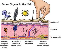
Sensation: Contains specialized sensory receptors for touch, pain, temperature, and pressure.
Excretion of Wastes: Sweat glands excrete metabolic wastes such as urea and salts.
Blood Reservoir: Stores 8–10% of the body’s total blood volume, which can be redirected as needed.
Vitamin D Synthesis: Initiates the synthesis of vitamin D upon exposure to UV light, essential for calcium absorption.
Regulation of Body Temperature: Sweat glands and blood vessels in the skin help maintain thermal homeostasis through evaporative cooling and vasodilation/vasoconstriction.

Structure of the Skin
Major Layers of the Skin
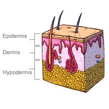
The skin is composed of three main layers, each with distinct structures and functions:
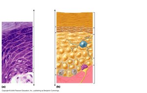
Epidermis: The outermost layer, composed of stratified squamous epithelium. It is replaced every 25–45 days and contains four main cell types.
Dermis: The middle layer, made of connective tissue, providing strength and elasticity. It contains blood vessels, nerves, and accessory structures.
Hypodermis (Subcutaneous Layer): Located beneath the dermis, primarily composed of adipose tissue, serving as insulation and energy storage.
Epidermal Cell Types
Keratinocytes: Produce keratin, a protein that provides strength and waterproofing to the skin.
Melanocytes: Synthesize melanin, the pigment responsible for skin color and UV protection.
Merkel Cells: Function as touch receptors in association with sensory nerve endings (Merkel discs).
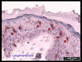
Langerhans’ Cells: Specialized macrophages that participate in immune defense; originate from bone marrow.
Layers of the Epidermis (Deep to Superficial)
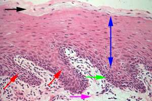
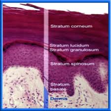
Stratum Basale: The deepest, mitotically active layer containing the youngest keratinocytes, melanocytes, and Merkel cells.
Stratum Spinosum: Known as the "prickly layer," contains keratinocytes and Langerhans’ cells.
Stratum Granulosum: The "granular layer" where cells flatten, accumulate keratin, and begin to lose nuclei and organelles.
Stratum Lucidum: A thin, clear layer found only in thick skin (e.g., palms, soles).
Stratum Corneum: The outermost "horny layer," consisting of dead, cornified cells; makes up up to ¾ of the epidermis’ thickness.
Dermis
Structure and Layers of the Dermis
The dermis is a strong, flexible connective tissue layer that supports and nourishes the epidermis. It is divided into two layers:
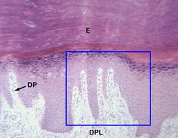
Papillary Layer: Composed of areolar connective tissue, contains dermal papillae (which form fingerprints) and Meissner’s corpuscles (touch receptors).
Reticular Layer: Made of dense irregular connective tissue, contains Pacinian corpuscles (pressure receptors), hair follicles, sebaceous (oil) glands, and sudoriferous (sweat) glands.
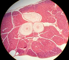
Hypodermis (Subcutaneous Layer)
The hypodermis lies just beneath the skin and is primarily composed of adipose tissue. It functions as insulation, shock absorption, and an energy reserve.
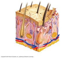
Homeostatic Imbalances
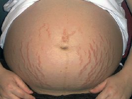
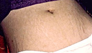
Stretch Marks (Striae)
Stretch marks are silvery-white scars that result from the tearing of the dermis due to rapid stretching, such as during pregnancy or significant weight changes. These marks persist even after the original cause (e.g., pregnancy) is resolved.
Summary Table: Layers and Cell Types of the Skin
Layer | Main Cell Types | Key Features |
|---|---|---|
Stratum Basale | Keratinocytes, Melanocytes, Merkel cells | Mitotically active, deepest layer |
Stratum Spinosum | Keratinocytes, Langerhans’ cells | Prickly appearance, immune defense |
Stratum Granulosum | Keratinocytes | Keratin accumulation, cell death begins |
Stratum Lucidum | Keratinocytes | Clear layer, only in thick skin |
Stratum Corneum | Dead keratinocytes | Thick, protective, outermost layer |
Additional info: The skin’s ability to regenerate, its role in immune defense, and its function as a sensory organ are critical for maintaining overall health and homeostasis.
