 Back
The Integumentary System: Structure, Function, and Clinical Relevance
Study Guide - Smart Notes
 Tailored notes based on your materials, expanded with key definitions, examples, and context.
The Integumentary System
Overview
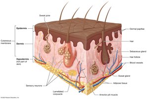
The integumentary system consists of the skin and its accessory structures, including hair, nails, and glands. It is the largest organ system in the body, accounting for 10-15% of total body weight. The skin is composed of two main layers: the superficial epidermis and the deeper dermis. Below the dermis lies the hypodermis, which is not considered part of the skin for this course.
Accessory structures: Nails, hair, sweat glands, sebaceous glands
Sensory neurons and receptors: Present in the skin
Arrector pili muscles: Attach to hair follicles
Epidermis: Avascular; superficial layers consist of dead cells
Dermis: Vascularized; provides nutrients and support
Functions of the Integumentary System
Vital Functions
The integumentary system is essential for maintaining homeostasis and protecting the body from external threats.
Protection: Shields underlying tissues from mechanical trauma, pathogens, and environmental threats. The skin's acid mantle deters pathogen growth.
Sensation: Sensory receptors detect harmful stimuli, aiding in homeostasis.
Thermoregulation: Maintains stable internal temperature via negative feedback loops.
Excretion: Eliminates small amounts of metabolic waste through sweat.
Synthesis of Vitamin D: Required for calcium absorption and bone health.
Thermoregulation
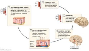
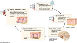
Thermoregulation is achieved through negative feedback loops that respond to changes in body temperature.
When body temperature rises: Thermoreceptors in the brain detect the change, the hypothalamus activates heat-loss mechanisms (sweating, vasodilation), and temperature returns to normal.
When body temperature falls: Thermoreceptors in the skin detect the change, the hypothalamus activates heat-conserving mechanisms (vasoconstriction), and temperature returns to normal.
Skin Structure
The Epidermis
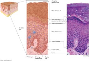
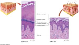
The epidermis is the superficial layer of the skin, composed primarily of keratinocytes and organized into five distinct strata.
Keratinocytes: Make up 95% of epidermal cells; produce keratin for strength and resistance to stress.
Stratum Basale: Deepest layer; mitotically active stem cells, melanocytes, and Merkel cells.
Stratum Spinosum: Thickest layer; metabolically active, contains dendritic cells.
Stratum Granulosum: Contains granules; forms water barrier and acid mantle.
Stratum Lucidum: Clear layer found only in thick skin.
Stratum Corneum: Several layers of dead, flattened keratinocytes; cells are shed regularly.
Thick and Thin Skin
Thick skin is found in areas subject to mechanical stress and contains all five strata, while thin skin covers the rest of the body and lacks the stratum lucidum.
Thick skin: Palms, soles, fingers, toes; no hair follicles, many sweat glands.
Thin skin: Most of the body; includes hair follicles, sweat, and sebaceous glands.
Callus: Formed by additional layers of stratum corneum in response to repeated pressure.
Other Cells of the Epidermis
Dendritic (Langerhans) cells: Immune phagocytes in the stratum spinosum.
Tactile (Merkel) cells: Sensory receptors in the stratum basale.
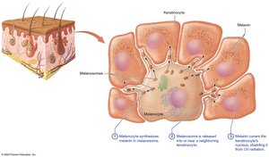
Melanocytes: Produce melanin in the stratum basale.
The Dermis
Structure and Layers
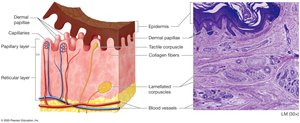
The dermis is the deep layer of the skin, providing structural support, blood supply, and housing sensory receptors. It is divided into two layers:
Papillary layer: Superficial; loose connective tissue, dermal papillae, Meissner corpuscles.
Reticular layer: Deep; dense irregular connective tissue, blood vessels, sweat glands, hair follicles, Pacinian corpuscles.
Skin Markings
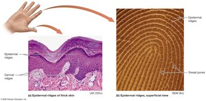
Dermal ridges: Form epidermal ridges, creating unique fingerprint patterns.
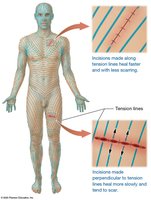
Tension (cleavage) lines: Gaps between collagen bundles; important for surgical incisions.
Flexure lines: Deep creases in areas such as the palms.
Skin Pigmentation
Melanin
Melanin is produced by melanocytes in the stratum basale and protects DNA from UV radiation. It is synthesized from tyrosine by the enzyme tyrosinase and transported to keratinocytes.
Eumelanin: Black and brown pigment
Pheomelanin: Yellow and red pigment
Melanin function: Shields DNA, must be replaced regularly
Other Pigments
Carotene: Yellow-orange pigment from diet; accumulates in stratum corneum
Hemoglobin: Red pigment in blood; gives skin a pinkish hue in fair-skinned individuals
Skin Color as a Diagnostic Tool
Erythema: Increased blood flow; red appearance
Pallor: Decreased blood flow; pale appearance
Cyanosis: Low oxygen; bluish appearance
Bilirubin: Accumulation causes jaundice; yellow-orange appearance
Accessory Structures: Hair, Nails, and Glands
Hair
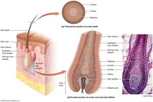
Hair is composed of keratinized epithelial cells and functions in protection and sensation. It consists of the shaft, root, and bulb, and grows in cycles.
Hair shaft: Dead keratinized cells; medulla, cortex, cuticle
Hair root: Embedded in dermis; living cells, matrix, follicle, arrector pili muscle
Hair bulb: Base of root; contains hair papilla with capillaries
Nails
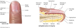
Nails are hard structures at the ends of digits, composed of stratified squamous epithelium and hard keratin. They protect fingertips and aid in manipulation.
Nail plate: Nail body (visible), nail root (under skin, matrix)
Protective folds: Proximal nail fold, eponychium (cuticle), medial/lateral folds, hyponychium
Lunula: Opaque half-moon region near proximal nail fold
Glands
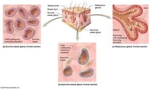
The skin contains several types of glands, each with distinct functions and modes of secretion.
Eccrine sweat glands: Most prevalent; thermoregulation; watery sweat
Apocrine sweat glands: Found in axillae, anal area, areolae; thick, protein-rich sweat; odor develops after bacterial metabolism
Ceruminous glands: Modified apocrine glands; produce cerumen (ear wax)
Mammary glands: Specialized sweat glands; produce milk
Sebaceous glands: Branched glands; produce sebum via holocrine secretion; associated with acne
Clinical Relevance: Skin Cancer
Types of Skin Cancer
Skin cancer is a common disease linked to UV exposure and other carcinogens. It results from mutations that cause uncontrolled cell growth.
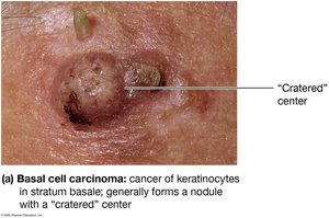
Basal cell carcinoma: Most common; arises from stratum basale keratinocytes; rarely metastasizes
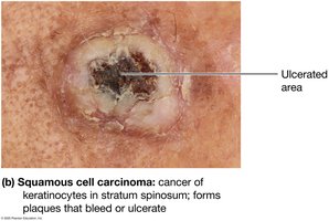
Squamous cell carcinoma: Second most common; arises from stratum spinosum keratinocytes; may metastasize
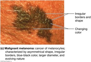
Malignant melanoma: Least common but most dangerous; arises from melanocytes; high tendency to metastasize
ABCDE Rule for Melanoma
A: Asymmetrical shape
B: Border irregularity
C: Color variation
D: Diameter > 6 mm
E: Evolving shape and size
Summary Table: Layers of the Skin
Layer | Location | Main Cell Types | Key Functions |
|---|---|---|---|
Stratum Basale | Deepest epidermal layer | Stem cells, melanocytes, Merkel cells | Cell division, pigment production, touch sensation |
Stratum Spinosum | Above basale | Keratinocytes, dendritic cells | Keratin production, immune defense |
Stratum Granulosum | Middle layer | Keratinocytes | Water barrier, acid mantle |
Stratum Lucidum | Thick skin only | Dead keratinocytes | Protection |
Stratum Corneum | Superficial layer | Dead keratinocytes | Barrier, prevents water loss |
Summary Table: Gland Types
Gland Type | Location | Secretion | Function |
|---|---|---|---|
Eccrine | Most skin areas | Watery sweat | Thermoregulation |
Apocrine | Axillae, anal, areolae | Protein-rich sweat | Odor after bacterial metabolism |
Ceruminous | Ear canal | Cerumen (ear wax) | Protects tympanic membrane |
Mammary | Breasts | Milk | Nourishes infants |
Sebaceous | Face, scalp | Sebum (oil) | Hydrophobic barrier, acne |
Additional info: Academic context was added to clarify the structure, function, and clinical relevance of the integumentary system, including expanded explanations of cell types, feedback mechanisms, and diagnostic features.
