 Back
The Integumentary System: Structure, Function, and Components
Study Guide - Smart Notes
 Tailored notes based on your materials, expanded with key definitions, examples, and context.
The Integumentary System
Overview
The integumentary system is composed of the skin and its derivatives, including hair, nails, sweat glands, and sebaceous (oil) glands. It serves as the body's primary barrier against the external environment and plays critical roles in protection, sensation, temperature regulation, and metabolic functions.
Skin: The largest organ of the body, consisting of multiple layers.
Hair: Provides protection and sensory input.
Nails: Protect the distal phalanx and enhance fine touch.
Sweat glands: Involved in thermoregulation and excretion.
Sebaceous glands: Secrete sebum for lubrication and protection.
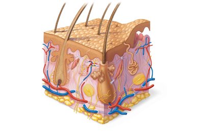
General Characteristics of the Skin
Skin Layers
The skin is composed of three main layers, each with distinct structures and functions:
Epidermis: The outermost layer, made of stratified squamous epithelial tissue; avascular.
Dermis: Middle layer, primarily fibrous connective tissue; vascular and contains nerves, glands, and hair follicles.
Hypodermis (Subcutaneous tissue): Not technically part of the skin; mostly adipose tissue that absorbs shock, insulates, and anchors skin to underlying structures (mainly muscles).
Epidermis
Cell Types
The epidermis contains several specialized cell types, each with unique functions:
Keratinocytes: Produce keratin, a fibrous protein that provides protective properties; most abundant cell type in the epidermis; millions are shed daily.
Melanocytes: Located in the deepest layer; produce melanin pigment, which is transferred to keratinocytes and protects the nucleus from UV damage.
Dendritic (Langerhans) cells: Star-shaped macrophages that patrol the epidermis and are part of the innate immune system.
Tactile (Merkel) cells: Sensory receptors for touch, associated with sensory nerve endings.
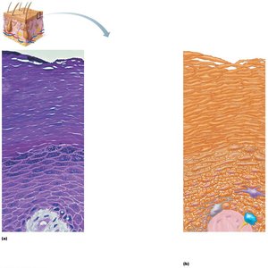
Layers of the Epidermis
The epidermis is organized into distinct layers (strata), each with specific characteristics:
Stratum basale (basal layer): Single row of mitotic stem cells; contains melanocytes and Merkel cells; cells migrate toward the surface, dying as they move upward (takes 25–45 days).
Stratum spinosum: Several layers thick; contains pre-keratin filaments, melanosomes, and dendritic cells.
Stratum granulosum: 1–5 layers of flattened cells; keratinization begins; cells accumulate granules and glycolipids, which slow water loss; cells above this layer die due to lack of nutrients.
Stratum lucidum: Present only in thick skin (e.g., palms, soles); thin, translucent band of dead keratinocytes.
Stratum corneum: 20–30 layers of flat, anucleated, keratinized dead cells; provides a durable overcoat for the body.
Dermis
Structure and Function
The dermis is a strong, flexible connective tissue layer beneath the epidermis. It contains fibroblasts, macrophages, nerves, blood vessels, lymphatics, hair follicles, and glands. The dermis is divided into two layers:
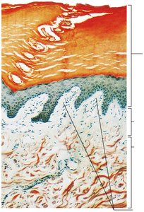
Papillary layer: Made of areolar connective tissue with interlacing collagen and elastic fibers; contains dermal papillae (finger-like projections) that house capillary loops, touch receptors, and pain receptors.
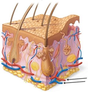
Reticular layer: Accounts for ~80% of dermal thickness; composed of dense irregular connective tissue with collagen and elastic fibers; provides strength, extensibility, and elasticity; contains the cutaneous plexus (vascular network). binds to water.
cutaneous plexua
-vascular
Skin Appendages
Hair and Hair Follicles
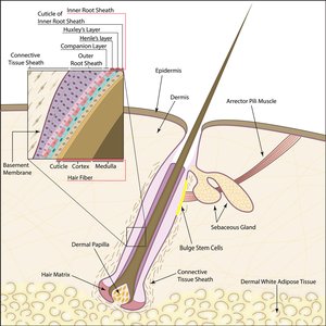
Hairs (pili) are flexible strands of dead, keratinized cells produced by hair follicles. Hair consists of hard keratin, which is more durable than the soft keratin found in the skin. Hair color is determined by melanins produced by melanocytes in the hair follicle.
Hair matrix: Actively dividing area of the bulb that produces hair cells.
Arrector pili: Smooth muscle attached to the follicle; responsible for "goose bumps."
Hair papilla: Dermal tissue containing capillaries that supply nutrients to growing hair.
Papillary Layer
Areolar connective tissue
interlacing collagen and elastic fibers
vascular
Phagocytes
Dermal papilla
finger like projections
Sweat Glands (Sudoriferous Glands)
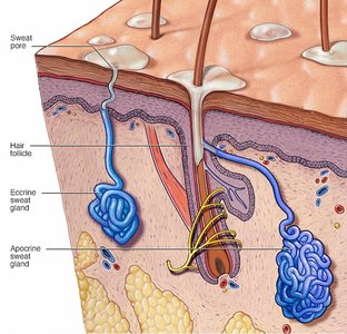
Sweat glands are distributed over most skin surfaces, with about 3 million per person. They are classified into two main types:
Eccrine (merocrine) sweat glands: Most numerous; secrete watery sweat for thermoregulation.
Apocrine sweat glands: Found in axillary and anogenital areas; secrete a thicker fluid; become active at puberty.
Ceruminous glands: Modified apocrine glands in the external ear canal; secrete cerumen (earwax).
exocrine ducts vs endocrine
Sweat glands contain myoepithelial cells that contract upon nervous system stimulation to force sweat into ducts.
Functions of the Skin
Protection
The skin acts as a barrier against microorganisms, abrasions, temperature extremes, and harmful chemicals. It provides:
Chemical barrier: Secretes chemicals (sweat, sebum) with low pH; melanin protects against UV radiation.
Physical barrier: Flat, dead, keratinized cells surrounded by glycolipids prevent water loss and entry of substances.
Biological barriers: Epidermal dendritic cells and dermal macrophages provide immune defense.
Body Temperature Regulation
Insensible perspiration: Unnoticeable sweat that occurs under normal conditions.
Sensible perspiration: Noticeable sweat produced to cool the body during heat or exercise.
In cold environments, dermal blood vessels constrict to reduce heat loss.
Cutaneous Sensations
The skin contains sensory receptors (exteroceptors) that respond to external stimuli such as temperature, touch, and pain. Free nerve endings detect painful stimuli.
Metabolic Functions
Synthesizes vitamin D when exposed to sunlight.
Keratinocytes can help disarm some carcinogens.
Excretion
Secretes limited amounts of nitrogenous wastes, including ammonia, urea, and uric acid.
