 Back
BackThe Integumentary System: Structure, Function, and Clinical Relevance
Study Guide - Smart Notes
 Tailored notes based on your materials, expanded with key definitions, examples, and context.
The Integumentary System
Overview of the Integumentary System
The integumentary system is composed of the skin and its accessory structures, including hair, nails, and glands. As the largest organ system, it plays a critical role in protection, sensation, thermoregulation, excretion, and vitamin D synthesis.
Skin (Cutaneous Membrane): Composed of two main layers: the superficial epidermis and the deeper dermis.
Accessory Structures: Include hair, nails, sweat glands, and sebaceous glands.
Hypodermis (Subcutaneous Tissue): Lies below the dermis, composed of loose connective and adipose tissue; not considered part of the skin but anchors it to underlying tissues.
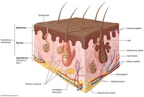
Skin Structure
Epidermis
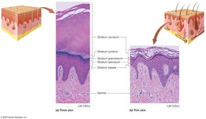
The epidermis is the outermost layer of the skin, consisting of keratinized stratified squamous epithelium. It is avascular and contains several distinct cell types and layers (strata).
Keratinocytes: Make up about 95% of epidermal cells; produce keratin for strength and water resistance.
Strata of the Epidermis (deep to superficial):
Stratum Basale (Germinativum): Single layer of stem cells; mitotically active; produces keratin and vitamin D precursors.
Stratum Spinosum: Thickest layer; spiny-shaped cells; active in keratin and vitamin D production.
Stratum Granulosum: 3-5 rows; contains keratin bundles and membrane-coating granules; forms water barrier.
Stratum Lucidum: Clear layer of dead keratinocytes; found only in thick skin.
Stratum Corneum: Multiple layers of dead, flattened keratinocytes; cells are shed regularly.
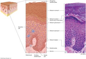
Mnemonic for Epidermal Layers
"Brilliant Studying Gives Loads of Confidence" for Basale, Spinosum, Granulosum, Lucidum, Corneum.
Keratinocyte Life Cycle
Keratinocytes originate in the stratum basale and are pushed upward through the strata, eventually being sloughed off in the stratum corneum. This process takes approximately 40-50 days.
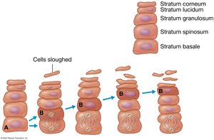
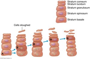
Other Epidermal Cells
Dendritic (Langerhans) Cells: Immune cells in the stratum spinosum; phagocytose pathogens.
Tactile (Merkel) Cells: Sensory receptors in the stratum basale; detect light touch.
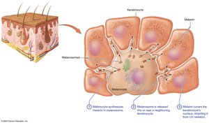
Melanocytes: Produce melanin pigment; found in the stratum basale.
Thick vs. Thin Skin
Thick Skin: Found on palms, soles, and palmar/plantar surfaces; contains all five strata; lacks hair follicles.
Thin Skin: Covers most of the body; lacks stratum lucidum; contains hair follicles and glands.
Callus: Thickened stratum corneum due to repeated pressure.
The Dermis
Structure and Function
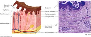
The dermis is the deep layer of the skin, providing structural support, housing blood vessels, sensory receptors, and anchoring the epidermis.
Papillary Layer: Superficial; loose connective tissue; forms dermal papillae that supply blood and house tactile corpuscles.
Reticular Layer: Deep; dense irregular connective tissue; contains blood vessels, glands, hair follicles, and lamellated corpuscles for deep pressure sensation.
Skin Markings
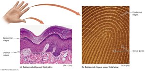
Dermal Ridges: Prominent in thick skin; form epidermal ridges (fingerprints).
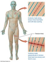
Tension (Cleavage) Lines: Gaps between collagen bundles; important for surgical incisions.
Flexure Lines: Deep creases in areas of joint movement (e.g., palms).
Skin Wrinkles
Wrinkles result from decreased collagen, elastic fibers, proteoglycans, and adipose tissue with age. UV exposure and smoking accelerate this process. Treatments include Botox, fillers, peels, and topical creams, though most nonprescription creams have limited effect.


Melanin and Skin Color
Melanin Production and Function
Melanin is produced by melanocytes in melanosomes and transferred to keratinocytes. It protects DNA from UV radiation but must be continually replaced as it degrades.
Tyrosinase: Enzyme that synthesizes melanin from tyrosine.
UV Exposure: Increases melanin production; immediate darkening and delayed DNA damage response.
Vitamin D Regulation: Melanin reduces vitamin D synthesis to maintain homeostasis.
Other Pigments Affecting Skin Color
Carotene: Yellow-orange pigment from diet; accumulates in stratum corneum.
Hemoglobin: Red pigment in blood; gives skin a pinkish hue in fair-skinned individuals.
Skin Color as a Diagnostic Tool
Erythema: Increased blood flow; red appearance.
Pallor: Decreased blood flow; pale appearance.
Cyanosis: Low oxygen; bluish color.
Jaundice: High bilirubin; yellow-orange color.
Accessory Structures of the Integument
Hair
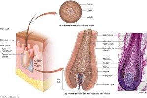
Hair (pili) is composed of keratinized epithelial cells and serves protective and sensory functions. It is found on most body surfaces except thick skin, lips, and parts of the genitalia.
Structure: Hair shaft (dead cells), root (living cells), follicle (epidermal and dermal sheaths), and bulb (base with papilla and matrix).
Growth: Alternates between growth and resting stages; influenced by genetics and hormones.
Pigment and Texture: Determined by melanin type and amount; changes with age.
Nails
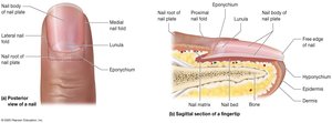
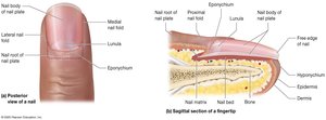
Nails are hard structures at the ends of digits, composed of hard keratin. They protect fingertips and aid in manipulation.
Structure: Nail plate (body and root), nail bed, matrix, folds, cuticle (eponychium), and lunula.
Growth: Continuous; fingernails grow faster than toenails.
Pigment: Nails are translucent; color reflects blood oxygenation.
Glands
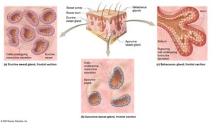
Eccrine Sweat Glands: Most common; secrete watery sweat for thermoregulation.
Apocrine Sweat Glands: Found in axillae, anal, and areolar regions; secrete protein-rich sweat onto hair follicles; odor develops when metabolized by bacteria.
Ceruminous Glands: Modified apocrine glands; produce ear wax (cerumen).
Mammary Glands: Specialized for milk production.
Sebaceous Glands: Secrete sebum (oily substance) via holocrine secretion; lubricates and waterproofs skin and hair.
Acne
Acne vulgaris is caused by the accumulation of sebum and dead cells in sebaceous glands, leading to comedones (blackheads) and, if infected, pustules (pimples). Hormones, especially testosterone, play a significant role.

Wounds, Burns, and Skin Cancer
Wounds
Wounds are disruptions in skin integrity, including lacerations, burns, and cancers.
Burns
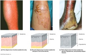
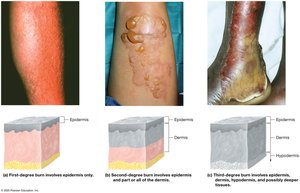
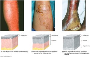
First-Degree: Only epidermis; redness and minor pain; no blisters.
Second-Degree: Epidermis and part/all of dermis; pain, blisters, possible scarring.
Third-Degree: Full thickness; damages all skin layers and possibly deeper tissues; risk of dehydration and infection; may require grafting.
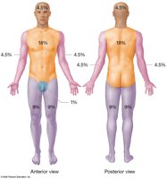
Rule of Nines
Used to estimate the percentage of body surface area affected by burns for fluid resuscitation. The body is divided into regions representing 9% (or multiples) of total surface area.
Skin Cancer
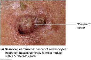
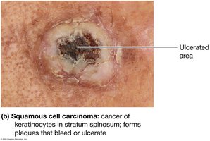
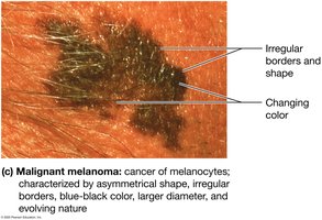
Skin cancer arises from mutations in skin cells, often due to UV exposure. The three main types are:
Basal Cell Carcinoma: Most common; arises from stratum basale; rarely metastasizes.
Squamous Cell Carcinoma: From stratum spinosum; may metastasize; forms scaly plaques.
Malignant Melanoma: Most dangerous; arises from melanocytes; high metastatic potential; identified by the ABCDE rule (Asymmetry, Border, Color, Diameter, Evolving).
Summary Table: Layers of the Epidermis
Stratum | Location | Key Features |
|---|---|---|
Basale | Deepest | Mitotically active stem cells, melanocytes, tactile cells |
Spinosum | Above basale | Thickest, keratin production, dendritic cells |
Granulosum | Middle | Keratin bundles, water barrier |
Lucidum | Thick skin only | Clear, dead keratinocytes |
Corneum | Superficial | Dead, flattened cells, keratinized, shed regularly |
Key Equations
Melanin Synthesis:
Clinical Applications
Skin color changes can indicate underlying health issues (e.g., cyanosis, jaundice).
Burn assessment is critical for fluid management and prognosis.
Early detection of skin cancer improves outcomes; use the ABCDE rule for melanoma screening.
