 Back
BackThe Integumentary System: Structure, Function, and Pathology
Study Guide - Smart Notes
 Tailored notes based on your materials, expanded with key definitions, examples, and context.
The Integumentary System
Overview of the Integumentary System
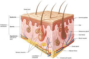
The integumentary system is the body's largest organ system, comprising the skin and its accessory structures. It plays a vital role in protection, sensation, thermoregulation, excretion, and vitamin D synthesis, all of which are essential for maintaining homeostasis.
Cutaneous membrane (skin): Consists of two main components:
Epidermis: The superficial layer, made of keratinized stratified squamous epithelium resting on a basement membrane.
Dermis: Located beneath the epidermis, composed of loose and dense irregular connective tissue.
Accessory structures: Sweat glands, sebaceous glands, hair, and nails are embedded within the skin.
Hypodermis: Also known as superficial fascia or subcutaneous fat, lies deep to the dermis and anchors the skin to underlying structures.
Functions of the Integumentary System
The integumentary system is essential for protecting underlying organs and maintaining homeostasis through several mechanisms:
Protection: The keratinized epithelium provides a durable barrier against mechanical trauma, pathogens, and environmental hazards. Immune cells (Langerhans cells) and antimicrobial secretions further protect against infection.
Sensation: Numerous sensory receptors in the skin detect changes in the internal and external environment, including heat, cold, and pain.
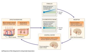
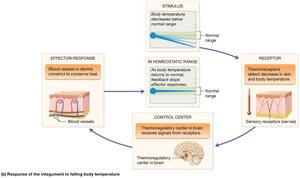
Thermoregulation: The skin regulates body temperature via negative feedback loops, involving sweating and vasodilation or vasoconstriction of dermal blood vessels.
Excretion: The skin eliminates small amounts of waste products and toxins.
Vitamin D synthesis: UV radiation converts cholesterol in the epidermis to cholecalciferol, which is then processed by the liver and kidneys to form calcitriol, essential for calcium absorption.
The Epidermis
Structure and Cell Types
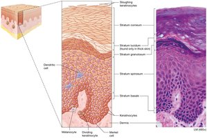
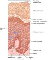
The epidermis is the most superficial layer of the skin, composed primarily of keratinocytes, which manufacture the tough protein keratin. Other cell types include dendritic (Langerhans) cells, Merkel cells, and melanocytes.
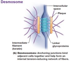
Keratinocytes: Make up about 95% of the epidermis; linked by desmosomes for structural integrity.
Dendritic (Langerhans) cells: Immune cells in the stratum spinosum.
Merkel cells: Sensory receptors in the stratum basale, detect light touch.
Melanocytes: Produce melanin, the pigment responsible for skin color.
Epidermal Layers (Strata)
The epidermis consists of five structurally distinct layers, from superficial to deep:
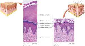
Stratum corneum: Outermost layer of dead, flattened keratinocytes.
Stratum lucidum: Clear layer found only in thick skin.
Stratum granulosum: Contains granules of keratin and lipid-based substances for waterproofing.
Stratum spinosum: Thickest layer, metabolically active.
Stratum basale: Single layer of stem cells, most mitotically active.
Keratinocyte Life Cycle
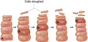
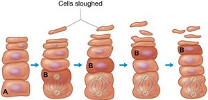
Keratinocytes are continuously replaced by mitosis in the stratum basale and spinosum, migrating through the layers and eventually being shed from the stratum corneum. This process takes 40–50 days.
Thick and Thin Skin
Thick skin (palms, soles) contains all five layers and a thick stratum corneum, but no hair follicles. Thin skin covers most of the body, has only four layers, and contains hair follicles, sweat glands, and sebaceous glands.
The Dermis
Structure and Layers
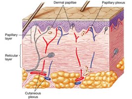
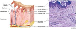
The dermis is a highly vascular layer beneath the epidermis, providing blood supply, sensory receptors, and anchoring the epidermis. It consists of two layers:
Papillary layer: Areolar connective tissue, contains dermal papillae and tactile (Meissner) corpuscles.
Reticular layer: Dense irregular connective tissue, contains collagen and elastic fibers, lamellated (Pacinian) corpuscles, blood vessels, glands, and adipose tissue.
Skin Markings
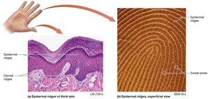
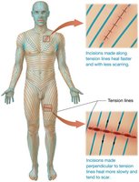
Skin markings such as dermal ridges, tension (cleavage) lines, and flexure lines are formed by the interaction between the dermis and epidermis. Dermal ridges create fingerprints, while tension lines indicate collagen fiber orientation.
Skin Pigmentation
Melanin
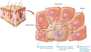
Melanin is produced by melanocytes in the stratum basale and protects keratinocyte DNA from UV-induced mutations. Its synthesis increases with UV exposure, resulting in tanning. Melanin also regulates vitamin D synthesis.


Common Variations in Pigmentation
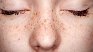
Freckles: Localized increased melanin production.
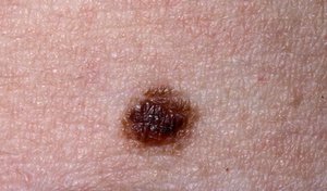
Moles (nevi): Local proliferation of melanocytes.
Albinism: Lack of tyrosinase, resulting in no melanin production.
Carotene and Hemoglobin
Carotene is a yellow-orange pigment from diet, accumulating in the stratum corneum. Hemoglobin, an iron-containing protein in red blood cells, gives skin a pinkish hue depending on blood flow in the dermis.
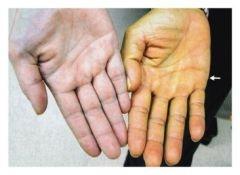
Skin Color as a Diagnostic Tool
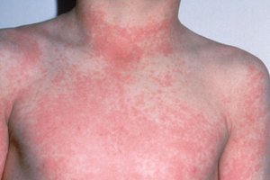
Erythema: Increased blood flow, causing redness.
Pallor: Decreased blood flow, causing paleness.
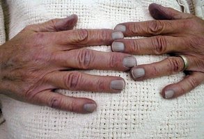
Cyanosis: Low oxygen levels, causing a bluish hue.

Accessory Structures: Hair, Nails, and Glands
Hair
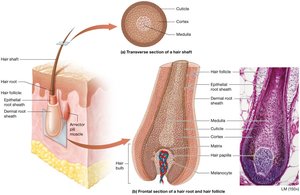
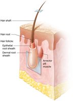
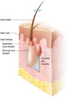
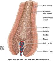
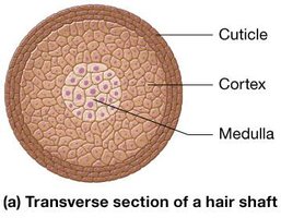
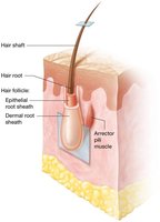
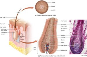
Hair is a filamentous structure derived from epithelium, consisting of a shaft and root. It provides protection, sensory input, and limited thermoregulation.
Shaft: Dead, keratinized cells projecting from the skin.
Root: Embedded in the dermis, surrounded by a hair follicle and sensory neurons.
Matrix: Actively dividing keratinocytes at the base of the root.
Arrector pili muscle: Smooth muscle causing piloerection (goosebumps).
Hair Pigment and Texture
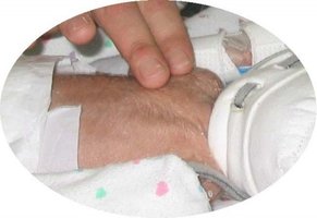
Hair color is determined by the type and amount of melanin produced by melanocytes in the matrix. Hair texture and type (lanugo, vellus, terminal) change with age and hormonal influences.




Nails
Nails are hard structures at the ends of digits, composed of stratified squamous epithelium filled with hard keratin. They protect underlying tissues and aid in gripping.
Nail plate: Visible part, divided into nail body and nail root.
Nail matrix: Site of active cell division.
Lunula: Half-moon shaped region of the nail plate.
Glands
The skin contains sweat (sudoriferous) and sebaceous glands, both derived from epidermal cells and located in the dermis.
Eccrine sweat glands: Most prevalent, secrete watery sweat for thermoregulation.
Apocrine sweat glands: Found in specific regions, secrete protein-rich fluid into hair follicles.
Ceruminous glands: Modified apocrine glands producing ear wax.
Mammary glands: Specialized sweat glands producing milk.
Sebaceous glands: Secrete oily sebum, providing a hydrophobic barrier and inhibiting bacterial growth.
Pathology of the Skin
Acne
Acne vulgaris is a common skin disorder caused by the accumulation of sebum and dead cells in sebaceous glands, often becoming infected and inflamed. Hormonal changes, especially testosterone, play a significant role.
Burns
Burns are classified by depth and extent:
First-degree: Damage to epidermis only; mild pain and redness.
Second-degree: Involves epidermis and part or all of dermis; pain, blistering, and scarring.
Third-degree: Involves epidermis, dermis, hypodermis, and possibly deeper tissues; severe damage, risk of dehydration and infection.
Rule of Nines
The rule of nines is a clinical tool for estimating the extent of burns, dividing the body into areas representing 9% of total body area.
Skin Cancer
Skin cancer is caused by mutations in DNA, often linked to UV exposure. The three main types are:
Basal cell carcinoma: Most common, arises from stratum basale, rarely metastasizes.
Squamous cell carcinoma: Arises from stratum spinosum, more likely to metastasize.
Malignant melanoma: Cancer of melanocytes, highly metastatic, diagnosed using the ABCDE rule.
ABCDE Rule for Melanoma Diagnosis
A: Asymmetrical shape
B: Border irregularity
C: Color variation
D: Diameter > 6 mm
E: Evolving shape and size
Additional info: The notes have been expanded with academic context, definitions, and examples to ensure completeness and clarity for college-level Anatomy & Physiology students.
